
- Title
-
N-Cadherin is essential for retinal lamination in the zebrafish
- Authors
- Erdmann, B., Kirsch, F.P., Rathjen, F.G., and Moré, M.I.
- Source
- Full text @ Dev. Dyn.
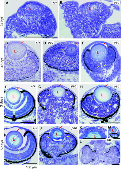
Eye development in the N-cadherin pacpaR2.10 mutant. A,B: Wild-type (WT) and mutant eyes in cross-section at 24 hours postfertilization (hpf). The WT eye cup is well formed and contains a lens (L) primordium and a pseudostratified retinal epithelium. Compared with the elongated and highly organized cells in the WT retina (arrowheads), mutant retinal cells are more disorganized and more rounded (arrowheads). C-E: WT and mutant eyes at 48 hpf. At this stage, the retinal lamination defect, including rosette formation, is clearly visible in the mutant (examples of rosettes are indicated by dotted lines). Arrows point to corresponding cell types in the WT and mutant: yellow arrows, retinal ganglion cells; white arrows, inner and outer plexiform layer; orange arrows, inner nuclear layer; green arrows, photoreceptors; red arrows, lens epithelium. The pigment epithelium can be easily seen because of its black pigment. E: In the mutant, it often does not fully surround the eye cup. Mutant cell bodies lack a uniform orientation like in WT. Unlike WT, the mutant optic nerve is often bordered by retinal ganglion cells (E, yellow arrows, compare with WT optic nerve in F). F-H: WT and mutant eyes of 3-day-old fish. Arrows are as above. In most mutants, eyes and brain are not fully separated (G, also J,L). The mutant lens epithelium in G (sectioned peripherally) contains some rounded-looking cells, whereas the mutant lens epithelium in H (sectioned centrally) is completely normal. I-M: WT and mutant eyes of 5-day-old fish. Arrows are as above. The lens is sometimes located off-center in the mutant, resulting in retinal sections without lens (L) and sections, where the lens (sectioned slightly peripherally) is not surrounded by much retina (M). Arrowheads in L indicate that, in the brain, rosettes are formed like in the retinae. The mutant photoreceptor cells appear more rounded and are sometimes moved away from the pigment epithelium, forming rosettes (green arrows). The mutant lens epithelium of the centrally sectioned lens in J shows clear abnormalities (blow up: K), in that cells are rounded and multilayered. |
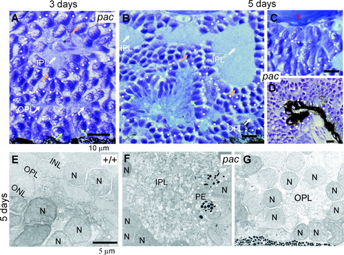
Rosette formation in the pac mutant. Arrows are as in Figure 1. A: Larger magnification of Figure 1D, showing three types of retinal rosettes at the 3-day stage: rosettes containing islands of inner plexiform layer tissue (IPL), rosettes containing islands of outer plexiform layer tissue (OPL), and rosettes of photoreceptors (green arrows). B: Larger magnification of Figure 1J, showing the same types of rosettes as in A at the 5-day stage. C: Cell cluster or rosette formed by retinal ganglion cells at day 5, magnified from Figure 1J. D: Rosette-like structures of the pigment epithelium, larger magnification of Figure 1L. E-G: Electron photomicrographs of 5-day-old retinae. E: WT outer plexiform layer and surrounding tissue. INL, inner nuclear layer; ONL, outer nuclear layer containing photoreceptor nuclei; N, nucleus. F: Mutant inner plexiform layer island and surrounding nuclei of the inner nuclear layer. Some mutant retinae contain small pigment epithelium (PE) islands within other retinal tissue. G: Mutant outer plexiform layer island, surrounded by nuclei of the inner or outer nuclear layer. Original magnifications = ×2,200 in E-G. |
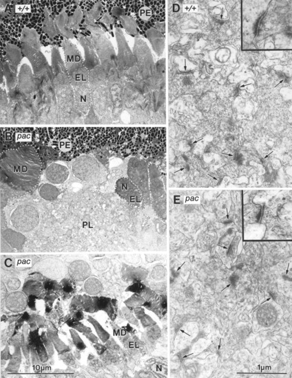
Ultrastructure of the eye region in 5-day-old wild-type (WT; A,D) and the N-cadherin pacpaR2.10 mutant (B,C,E) zebrafish. A: WT photoreceptor cells (example outlined) form membranous discs (MD) in the outer segment, partially surrounded by the pigment epithelium (PE). The inner segment or ellipsoid (EL) contains numerous mitochondria, and the nuclei (N) form the outer nuclear layer. B: The electron photomicrograph depicts a disturbed area of the outer mutant retina, including part of the pigment epithelium. In the mutant, photoreceptor cells are often not properly arranged in parallel like in WT. Photoreceptor orientation is sometimes even inverted, so that the nucleus lies closer to the pigment epithelium than the rest of the cell (cell labeled with N and EL; the MD segment is not in plane with the section). The (outer) plexiform layer (PL) comes close to the pigment epithelium, only separated by one row of nuclei. The mutant pigment epithelium cells are indistinguishable from WT. C: This electron photomicrograph depicts part of a rosette formed by photoreceptors similar to the one shown in Figure 2B. Note that although some photoreceptor cells are deformed (see also leftmost photoreceptor in B), a membranous disc segment and a segment containing mitochondria are formed like in WT. D: WT inner plexiform layer. E: Mutant inner plexiform layer island (of a rosette like shown in Fig. 2F). The number and morphology of synapses (arrows and insets) is indistinguishable between WT and mutant. Original magnifications = ×4,000 in A-C, ×30,000 in D,E, ×56,000 in insets. PHENOTYPE:
|

Unillustrated author statements PHENOTYPE:
|