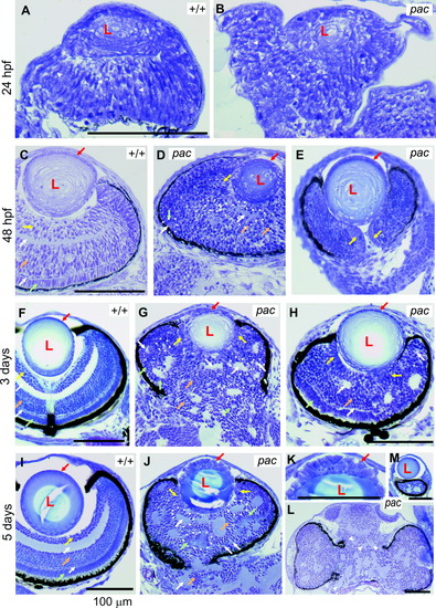
Eye development in the N-cadherin pacpaR2.10 mutant. A,B: Wild-type (WT) and mutant eyes in cross-section at 24 hours postfertilization (hpf). The WT eye cup is well formed and contains a lens (L) primordium and a pseudostratified retinal epithelium. Compared with the elongated and highly organized cells in the WT retina (arrowheads), mutant retinal cells are more disorganized and more rounded (arrowheads). C-E: WT and mutant eyes at 48 hpf. At this stage, the retinal lamination defect, including rosette formation, is clearly visible in the mutant (examples of rosettes are indicated by dotted lines). Arrows point to corresponding cell types in the WT and mutant: yellow arrows, retinal ganglion cells; white arrows, inner and outer plexiform layer; orange arrows, inner nuclear layer; green arrows, photoreceptors; red arrows, lens epithelium. The pigment epithelium can be easily seen because of its black pigment. E: In the mutant, it often does not fully surround the eye cup. Mutant cell bodies lack a uniform orientation like in WT. Unlike WT, the mutant optic nerve is often bordered by retinal ganglion cells (E, yellow arrows, compare with WT optic nerve in F). F-H: WT and mutant eyes of 3-day-old fish. Arrows are as above. In most mutants, eyes and brain are not fully separated (G, also J,L). The mutant lens epithelium in G (sectioned peripherally) contains some rounded-looking cells, whereas the mutant lens epithelium in H (sectioned centrally) is completely normal. I-M: WT and mutant eyes of 5-day-old fish. Arrows are as above. The lens is sometimes located off-center in the mutant, resulting in retinal sections without lens (L) and sections, where the lens (sectioned slightly peripherally) is not surrounded by much retina (M). Arrowheads in L indicate that, in the brain, rosettes are formed like in the retinae. The mutant photoreceptor cells appear more rounded and are sometimes moved away from the pigment epithelium, forming rosettes (green arrows). The mutant lens epithelium of the centrally sectioned lens in J shows clear abnormalities (blow up: K), in that cells are rounded and multilayered.
|
