|
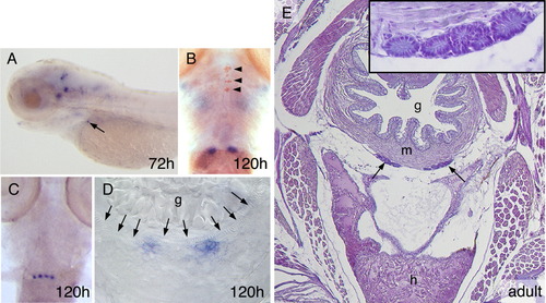
calca is expressed in the ultimobranchial bodies of zebrafish. A-D: calca expression (blue) in zebrafish larvae. Stages, bottom right. A: Expression in the brain and in the ultimobranchial bodies (arrow), lateral view, anterior to the left. B: Double staining with thyroglobulin (TG) immunostaining (arrowheads). C: A specimen with four ultimobranchial bodies instead of the usual two as in B. B and C are ventral views, anterior to the top. D: Cross-section on the level of the ultimobranchial bodies, note the muscle layer (arrows) around the gut that is still filled with yolk. E: Position of the ultimobranchial bodies in the adult zebrafish. Hematoxylin and eosin staining, section on the level of the sinus venosus of the heart. Inset: A close up of the ultimobranchial bodies visualizes their glandular appearance and organization as follicular epithelia. g, gut; h, heart; m, muscle layer of the gut.
|