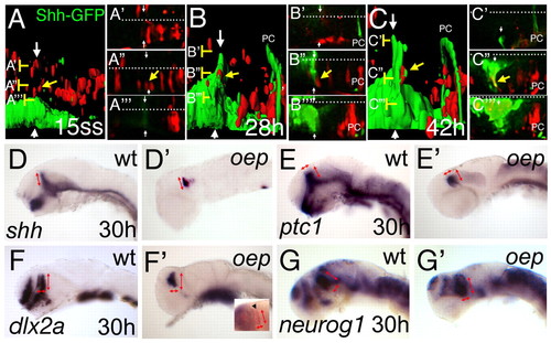
The basal plate is dispensable for ZLI formation and function. Rhodamine-dextran labelled cells (red) from a Shh-GFP transgenic embryo have been grafted into an embryo with the same genetic GFP-background (green) at sphere stage and further examined from 15 somites to 42 hpf (24 hours) on a confocal microscope. (A-C) Lateral views of the MDT of the same embryo at indicated stages after 3D reconstruction and surface rendering. (A'-A''', B'-B''', C'-C''') High magnification of the original dorsal sections of the scan at the indicated levels (brackets). White arrows indicate the ZLI and white dots indicate the midline. An indicated cell (yellow arrow) lies dorsal to the ZLI (A,A''). At 28 hpf, the cells are adjacent to the GFP positive ZLI (B,B'') and contribute to the ZLI at 42 hpf, shown by interdigitated red and green surfaces (C), and by a yellow overlay of the red rhodamine label with the GFP expression in the corresponding section (C''). PC marks the posterior commissure, marked by some shh-GFP positive axons. At 30 hours, oep mutant embryos were stained by indicated marker to analyse formation and function of the ZLI in embryos lacking the basal plate. In wild-type embryos, shh is expressed in the ventral neural tube as well as the ZLI (D). In oep mutants, shh can be detected only in the ZLI and the ventral hindbrain (D'). The dorsoventral extend of the ZLI is similar in wild-type and oep mutants (red arrows). ptc1 has a similar width to that of wild-type siblings in the diencephalon (E,E'; red arrows). dlx2a expression in the prethalamus has the same anteroposterior as well as dorsoventral extend in wild-type siblings as in oep mutants (F,F'; red arrows), as does neurog1 expression in the thalamus (G,G'; red arrows). Double in situ hybridization for the telencephalic marker emx1 in blue and dlx2a in red distinguishes the diencephalic dlx2a expression domain (inset in F', arrowhead).
|
