
Fig. 5
- ID
- ZDB-FIG-060112-22
- Publication
- Paulus et al., 2006 - Zebrafish bashful/laminin-alpha1 mutants exhibit multiple axon guidance defects
- Other Figures
- All Figure Page
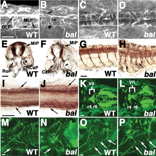
- Back to All Figure Page
Spinal and branchial axon phenotypes in bal. All views are anterior left except E,F (dorsal up). Labeling is anti-α-tubulin (A-D), ZNP-1 (E-H), F59 (I,J), or Tg(isl1:GFP; K-P). A-D, K-P are confocal projections. A-H: Embryos are wild-type (WT; A,C,E,G) or bal (B,D,F,H). A-D: Lateral (A,B) or dorsal (C,D) views of spinal cord in 23 hours postfertilization (hpf) embryos. dorsal longitudinal fasciculus (DLF; arrow in B) and primary commissural ascending neuron (CoPA) axons (arrowhead in B) are disorganized and the medial longitudinal fasciculus (MLF) is absent (asterisk in B) in bal (B). CoPA axons (arrows in C,D) correctly cross the midline. E-H: Cross-sections (E,F) or lateral views (G,H) of 36 hpf embryos showing extra axon running parallel to caudal primary axon (CaP; arrowhead in F), absence of normal rostral primary axon (RoP; open arrowhead in F) and axon branching (arrows in H). The asterisk in E indicates out of focus CaP in the next segment. I,J: Dorsal views of 22 hpf embryos showing normal slow muscle differentiation. K,L: Ventral views of hindbrain in 3 days postfertilization (dpf) embryos showing that some nVII branchiomotor neurons fail to migrate. In L, more nVII cells are in r4, rather than r6. M-P: Ventral (M,N) and lateral views (O,P) of branchiomotor axons innervating pharyngeal arches (arrows) in 4 dpf embryos. Scale bars = 20 μm in A-D, 40 μm in E-P. |