
- Title
-
Zebrafish bashful/laminin-alpha1 mutants exhibit multiple axon guidance defects
- Authors
- Paulus, J.D., and Halloran, M.C.
- Source
- Full text @ Dev. Dyn.
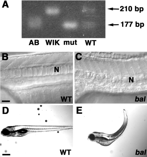
Mapping and morphology of baluw1 embryos. A: Gel of polymerase chain reaction products showing a band at 177 bp from AB founder (AB), baluw1 mutant embryos (mut), and wild-type sibling embryos (WT). The band at 210 bp is only present in WIK founder (WIK) and WT lanes. B,C: Lateral views (anterior left) of 24 hpf WT (B) and baluw1 (C) trunks. N denotes notochord. D,E: 5 days postfertilization (dpf) WT (D) and baluw1 (E) embryos. Scale bars = 20 μm in A,B, 200 μm in C,D. |

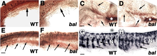
Retinal ganglion cell (RGC) axon defects in bal embryos. K-N are in situ hybridizations. All views are anterior left. A,B: Ventral views of ZN-5-labeled RGC axons in 2 days postfertilization (dpf) wild-type (WT; A) and bal (B) embryos showing ipsilateral projections in bal (arrows in B). C-E: Ventral views of 2.5 dpf WT (C) or bal (D,E) embryos injected with 1,1′-dioctadecyl-3,3,3′ ,3′-tetramethylindocarbocyanine perchlorate (DiI) into eye on bottom. The arrow in D indicates an ipsilateral projection, and the arrowhead in E shows the split optic tract. The dashed line indicates the midline. F,G: Lateral views, contralateral to injected eye, of 4 dpf WT (F) and bal (G) embryos injected with DiI (red) in nasal and DiA (green) in temporal retina. RGC axons extend anteriorly in bal (arrowhead). Injection sites of DiI and 4-4-dihexadecylaminostyryl-N-methylpyridinium iodide (DiA) overlap in G. H-J: Dorsal views of 5 dpf WT (H) and bal (I,J) embryos injected with DiI nasal and DiA temporal. Arrowheads indicate anterior projections, and the arrow denotes ipsilateral tectal projections. K,L: Lateral views of 48 hpf WT (K) and bal (L) embryos showing ephrinA5a in the retina (arrow). M,N: Ventral views of 48 hpf WT (M) and bal (N) embryos showing foxa2 along optic pathway (arrowheads) and in midbrain (arrows). E denotes eye. Scale bars = 60 μm. EXPRESSION / LABELING:
|

Retinal ganglion cell (RGC) axon defects in bal embryos. K-N are in situ hybridizations. All views are anterior left. A,B: Ventral views of ZN-5-labeled RGC axons in 2 days postfertilization (dpf) wild-type (WT; A) and bal (B) embryos showing ipsilateral projections in bal (arrows in B). C-E: Ventral views of 2.5 dpf WT (C) or bal (D,E) embryos injected with 1,1′-dioctadecyl-3, 3,3′,3′-tetramethylindocarbocyanine perchlorate (DiI) into eye on bottom. The arrow in D indicates an ipsilateral projection, and the arrowhead in E shows the split optic tract. The dashed line indicates the midline. F,G: Lateral views, contralateral to injected eye, of 4 dpf WT (F) and bal (G) embryos injected with DiI (red) in nasal and DiA (green) in temporal retina. RGC axons extend anteriorly in bal (arrowhead). Injection sites of DiI and 4-4-dihexadecylaminostyryl-N-methylpyridinium iodide (DiA) overlap in G. H-J: Dorsal views of 5 dpf WT (H) and bal (I,J) embryos injected with DiI nasal and DiA temporal. Arrowheads indicate anterior projections, and the arrow denotes ipsilateral tectal projections. K,L: Lateral views of 48 hpf WT (K) and bal (L) embryos showing ephrinA5a in the retina (arrow). M,N: Ventral views of 48 hpf WT (M) and bal (N) embryos showing foxa2 along optic pathway (arrowheads) and in midbrain (arrows). E denotes eye. Scale bars = 60 μm. |
bal embryos have defects in midbrain and hindbrain neurons. All are ventral views with anterior left. A-D: Nuclei of the medial longitudinal fasciculus (nucMLF) neurons labeled with ZN-12 in wild-type (WT; A,C) and bal (B,D) embryos at 24 hours postfertilization (hpf). C,D: Higher magnifications images of A,B. B: Cell bodies are laterally misplaced and disorganized in bal (arrow). B,D: Axons initially project in multiple directions (arrowheads in B,D) and then form a loose fascicle (asterisk in B). D: Arrow and inset shows cell body with multiple processes. E,F: Hindbrain neurons labeled with anti-α-tubulin in 24 hpf WT (E) and bal (F) embryos. Axons are defasciculated (asterisk), are found at the midline (arrows), or extend longitudinally along the midline (arrowhead). The open arrowhead shows the posterior most extent of axons in MLF. G-I: Mauthner neurons labeled with 3A10 in 27 hpf WT (G) and bal (H,I) embryos showing extra projection from soma (asterisks in I), anteriorly extending axons (arrow in I), branched axons (arrow in I), straight contralateral axon projection (arrow in H), posterior extent of axons (arrowheads in H,I). Scale bars = 40 μm for all full panels, 20 μm for inset in D. |
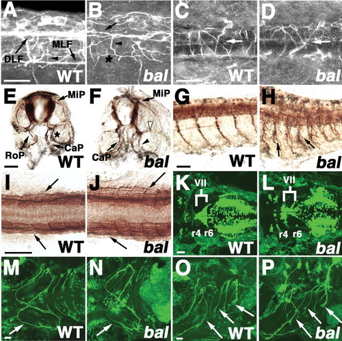
Spinal and branchial axon phenotypes in bal. All views are anterior left except E,F (dorsal up). Labeling is anti-α-tubulin (A-D), ZNP-1 (E-H), F59 (I,J), or Tg(isl1:GFP; K-P). A-D, K-P are confocal projections. A-H: Embryos are wild-type (WT; A,C,E,G) or bal (B,D,F,H). A-D: Lateral (A,B) or dorsal (C,D) views of spinal cord in 23 hours postfertilization (hpf) embryos. dorsal longitudinal fasciculus (DLF; arrow in B) and primary commissural ascending neuron (CoPA) axons (arrowhead in B) are disorganized and the medial longitudinal fasciculus (MLF) is absent (asterisk in B) in bal (B). CoPA axons (arrows in C,D) correctly cross the midline. E-H: Cross-sections (E,F) or lateral views (G,H) of 36 hpf embryos showing extra axon running parallel to caudal primary axon (CaP; arrowhead in F), absence of normal rostral primary axon (RoP; open arrowhead in F) and axon branching (arrows in H). The asterisk in E indicates out of focus CaP in the next segment. I,J: Dorsal views of 22 hpf embryos showing normal slow muscle differentiation. K,L: Ventral views of hindbrain in 3 days postfertilization (dpf) embryos showing that some nVII branchiomotor neurons fail to migrate. In L, more nVII cells are in r4, rather than r6. M-P: Ventral (M,N) and lateral views (O,P) of branchiomotor axons innervating pharyngeal arches (arrows) in 4 dpf embryos. Scale bars = 20 μm in A-D, 40 μm in E-P. |
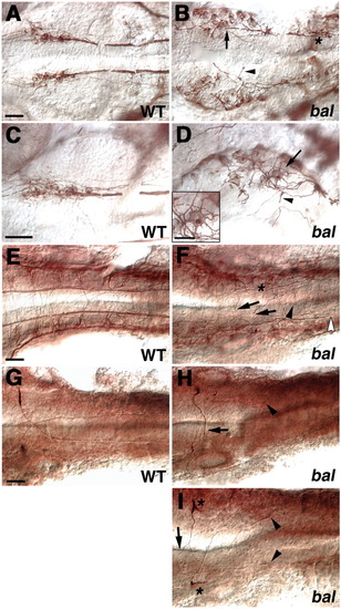
Other peripheral axons and migrating neural crest cells are normal in bal. A-F: Neurons were labeled with anti-α-tubulin (A,B), or ZN-12 (C-F). All are lateral views with anterior to the left. A,B: The 24 hours postfertilization (hpf) wild-type (WT; A) and bal (B) trunks showing posterior lateral line ganglion (PLLg) axons at same somite level (arrows). C,D: The 24 hpf WT (C) and bal (D) heads showing trigeminal axons extending from the trigeminal nucleus (asterisk) along the surface of the head (arrows). E,F: The 24 hpf WT (E) and bal (F) trunks showing Rohon-Beard axons extending along the surface of the trunk (arrows). G,H: In situ hybridization with crestin in 25 hpf WT (G) and bal (H) trunks showing ventral streams of neural crest cells (arrows). N denotes notochord. Scale bars = 40 μm. |

Unillustrated author statements |