
Fig. 4
- ID
- ZDB-FIG-060112-21
- Publication
- Paulus et al., 2006 - Zebrafish bashful/laminin-alpha1 mutants exhibit multiple axon guidance defects
- Other Figures
- All Figure Page
- Back to All Figure Page
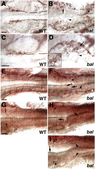
bal embryos have defects in midbrain and hindbrain neurons. All are ventral views with anterior left. A-D: Nuclei of the medial longitudinal fasciculus (nucMLF) neurons labeled with ZN-12 in wild-type (WT; A,C) and bal (B,D) embryos at 24 hours postfertilization (hpf). C,D: Higher magnifications images of A,B. B: Cell bodies are laterally misplaced and disorganized in bal (arrow). B,D: Axons initially project in multiple directions (arrowheads in B,D) and then form a loose fascicle (asterisk in B). D: Arrow and inset shows cell body with multiple processes. E,F: Hindbrain neurons labeled with anti-α-tubulin in 24 hpf WT (E) and bal (F) embryos. Axons are defasciculated (asterisk), are found at the midline (arrows), or extend longitudinally along the midline (arrowhead). The open arrowhead shows the posterior most extent of axons in MLF. G-I: Mauthner neurons labeled with 3A10 in 27 hpf WT (G) and bal (H,I) embryos showing extra projection from soma (asterisks in I), anteriorly extending axons (arrow in I), branched axons (arrow in I), straight contralateral axon projection (arrow in H), posterior extent of axons (arrowheads in H,I). Scale bars = 40 μm for all full panels, 20 μm for inset in D. |