
Fig. 6
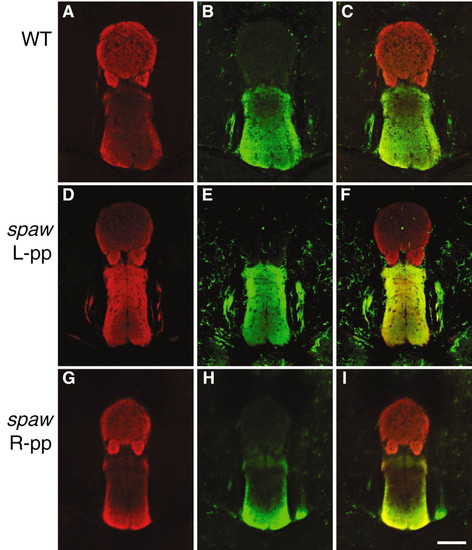
DV difference in Lov+ and Ron+ habenular projections onto the adult IPN. Confocal images of anti-Lov (red) and anti-Ron (green) double-labeled horizontal sections through adult midbrain of (A-C) WT or (D-F) left-positioned parapineal and (G-I) right-positioned parapineal fish selected from spaw MO-injected Tg(foxD3:GFP) embryos and reared separately. Vibratome sections (150 µm) vary slightly in their coordinates along anteroposterior axis of the brain, accounting for differences in immunofluorescent signals. Although intensity of labeling varies between given sections, DV regionalization of immunoreactive habenular axons at the IPN is highly stereotypic. Dorsal is at the top in all images. Scale bar: 60µm. |