
Fig. 4
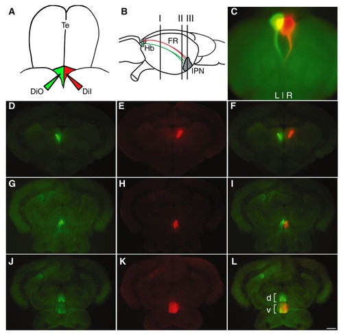
Selective labeling of L-R habenulae in adult zebrafish brain. (A) Schematic of adult zebrafish dorsal brain. The paired habenular nuclei lie at the dorsal surface of the diencephalon posterior to the telencephalon (Te) and anterior to the optic tectum. The habenular commissure was severed and DiI was injected into the right habenula (red) and DiO into the left habenula (green) (Aizawa et al., 2005). (B) Schematic of adult habenulointerpeduncular tract (sagittal view, anterior left) indicating approximate positions of transverse sections (150 µm) in D-L: I, D-F; II, G-I and III, J-L. (C) DiO- and (D-L) DiI-labeled left and right habenular projections along the fasciculus retroflexus to IPN viewed dorsally. Fluorescent images were captured using GFP3 (D,G,J) or rhodamine filter sets (E,H,K). (F,I,L) Digital overlay of DiO and DiI images. Dorsal (d) and ventral (v) regions of the target are indicated in L. Scale bar: 120 µm. |