|
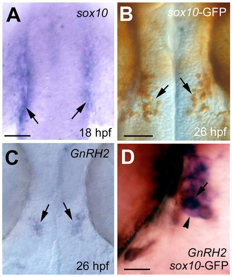
Co-localization of sox10-GFP and gnrh2 expression in the developing midbrain. (A) The expression sox10 is maintained in a small cluster of cells (arrows) at the backside of the developing optic cup at 18 hpf. (B) The GFP protein, recognized with an anti-GFP antibody at 26 hpf (arrows), reflects the earlier expression of sox10. (C) Differentiating gnrh2 cells (arrows) are found in the same region as the sox10-GFP immunoreactivity (see B). (D) gnrh2-expressing cells (purple, arrowhead) lie in the lateral neural tube, and in a subset of these cells the in situ signal (purple) co-localizes with the sox10-GFP immunoreactivity (purple+brown, arrowhead). Scale bars: A, 100 µm; B,C, 100 µm; D, 20 µm.
|