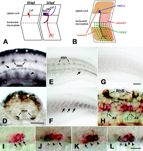
Expression of neuropilin-1a (NRP1a) and semaphorin 3A2 (sema3A2) in the trunk of embryonic zebrafish. A: A schematic side view of trunk segments at 18 and 24 hours postfertilization (hpf) is given. At 18 hpf, the caudal primary motor neuron (CaP) grows an axon out of the spinal cord. At 24 hpf, the axons of the middle (MiP) and rostral (RoP) primary motor neurons have followed on the common pathway to the horizontal myoseptum. The CaP axon is the only one growing ventrally beyond the horizontal myoseptum. B: Expression patterns of NRP1a (blue) and its potential ligands sema3A1 (orange), sema3A2 (red), and VEGF (green) are summarized for CaP in one trunk hemisegment. C: In a lateral view of a whole-mounted 16 hpf embryo at mid-trunk level (rostral is left), NRP1a mRNA is expressed in the dorsal spinal cord (asterisks), in motor neurons (mn), and in the hypochord (arrow). Arrowhead indicates expression in putative angioblasts. D: In a cross-section through the trunk at 16 hpf, expression is obvious in the dorsal spinal cord (asterisks) and the motor neurons (mn). E: In a lateral view of the caudal trunk of a 24 hpf embryo, expression of NRP1a is reduced in the dorsal spinal cord but is still strong in motor neurons (mn) and in a forming blood vessel (arrow). F: Sema3A2 is expressed in the caudal half of trunk myotomes (arrows; age and orientation as in E). G: In situ hybridization with an NRP1a sense RNA probe did not yield a signal (age and orientation as in E). H: A lateral view of a whole-mounted 24 hpf embryo is shown at caudal trunk level. NRP1a mRNA is labeled in red and tubulin protein in brown. Anti-tubulin-immunopositive motor axons (ax) can be seen to emerge from NRP1a mRNA-labeled motor neurons (mn). Rohon-Beard neurons (RhB) are labeled by the anti-tubulin antibody but not by the NRP1a in situ hybridization probe. I-L: Double labeling of NRP1a (red) and islet-1/-2 (brown) mRNAs at 24 hpf is shown (lateral trunk views, rostral is left). In I, the islet probes were omitted. In J, the NRP1a probe was combined with the islet-1 probe and in K with the islet-2 probe. In L, the NRP1a probe was combined with both probes. Arrows indicate cells labeled in red only (NRP1a). Arrowheads indicate double-labeled cells, and open arrowheads indicate cells labeled only in brown (islets). Scale bars = 25 μm in C,D,H, 50 μm in G (applies to E-G), 12.5 μm in L (applies to I-L).
|
