
Fig. 5
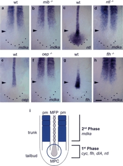
mdka transcription is not regulated by early acting floor plate-inducing factors. (a,b) In mib-/- embryos, identified by an enlarged notochord and an altered somite shape, mdka expression is normal compared with wild-type embryos (n = 14). (c,e,g) ntl, oep, and flh are expressed in the notochord of the trunk and progenitor cells in the tailbud. (c) In addition, ntl is also expressed in the posterior tailbud region. (d) In ntl-/- mutants that lack a notochord and have an enlarged MFP, mdka expression is not impaired (n = 24). In these embryos, the paraxial mesoderm is separated at the midline by an expanded MFP. (f) oep-/- mutants identified by their characteristic head phenotype form a normal notochord, but lack MFP. In oep-/-, mdka expression is normal (n = 11). (h) In flh-/- mutants, mdka is expressed at the midline (n = 25). (i) Two-phase model for MFP formation during neurulation in the zebrafish embryo. Midline precursor cells (MPC) in the tailbud are initially induced to adopt MFP fate in a first Mdka-independent phase. As the embryo extends caudally, Mdka derived from the trunk paraxial mesoderm (pm) regulates allocation of MFP cells (second phase). All pictures are dorsal views of the posterior trunk and tail; anterior is to the top. Arrowheads mark the position of the posterior front of mdka expression in the paraxial mesoderm. Bar: h, 100 µm.
|
| Genes: | |
|---|---|
| Fish: | |
| Anatomical Terms: | |
| Stage: | 10-13 somites |