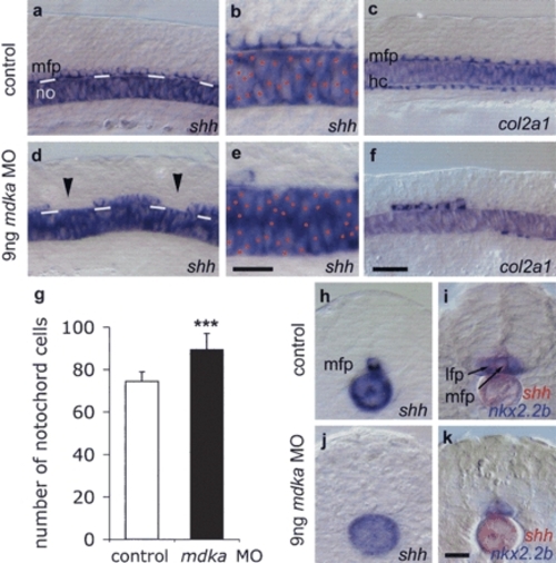
MFP formation requires Mdka protein. Lateral views of noninjected control (a-c) and 9 ng mdka MO morpholino-injected 12s embryos (d-f). mdka MO-injected embryos show gaps in the MFP (arrowheads; reduced MFP expression of shh in n = 103/136, 76% of injected embryos [d,e]; col2a1 in n = 114/188, 61% [f]; and f-spondin in n = 19/30, 63% [data not shown]) and increased cell density in the notochord (expressing shh [d,e] and ntl, n = 17/27, 63% [data not shown]). White lines in a and d indicate the border between the notochord and MFP. Red dots mark notochord cells in b and e. Large gaps were also observed in the hypochord (col2a1 reduced in n = 146/188, 78% of embryos [f]). (g) Mdka MO significantly increased the number of notochords cells: n = 10; (***) p < 0.001 versus control; Mann-Whitney U-test (see Supplementary Fig. S5). (h-k) Cross-sections of control (h,i) and mdka MO-injected (j,k) embryos at the trunk level showing loss of shh-positive MFP cells and fusion of nkx2.2b-expressing lateral floor plate cells (in n = 13/22, 59% of embryos). In several cases (n = 9/22, 41%) also lateral floor plate cells were missing, possibly due to insufficient levels of Shh. Bar: f, 20 μm; b,k, 10 μm. (hc) hypochord.
|
