
- Title
-
Expression of zebrafish rag genes during early development identifies the thymus
- Authors
- Willett, C.E., Zapata, A.G., Hopkins, N.A., and Steiner, L.A.
- Source
- Full text @ Dev. Biol.
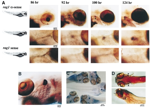
Whole-mount in situ hybridization of rag expression during zebrafish development. (A) Embryos and larvae were harvested at 3- hr intervals and hybridized with either a rag1 sense or antisense probe. Staining with the rag1 antisense probe shows expression in bilaterally symmetric spherical regions posterior to the eye. Top row, original magnification 55x; middle row, original magnification 110x; bottom row, staining with the rag1 sense control (original magnification 110x). (B) rag1 whole-mount in situ hybridization of a 2- week-old fish. (C) rag1 staining of several 1-week-old fish. (D)A 7-day-old fish hybridized to rag2 under the same whole-mount hybridization conditions used for rag1. EXPRESSION / LABELING:
|

Sections of fish hybridized to rag1 probe identify the zebrafish thymus. (A) A dorsal view of a 1-week-old fish hybridized with the rag1 antisense probe. The thymus (t) is clearly visible. A line across the posterior head indicates the plane of the section shown in B. The section in B has been stained with toluidine blue and shows the location of typically lymphoid organs in the head identified as the bilaterally symmetric thymus (t). The thymus lies just ventral to the otic vesicle (o) in the plane of the heart (h). C and D are sequential sections of the fish shown in A; in addition, C has been stained with toluidine blue which shows that this organ is filled with small cells possessing large nuclei, typical of thymocytes. Comparison of B, C, and D shows that the organ having a typically lymphoid appearance in B hybridizes to the rag1 probe. b, brain; e, eye; ga, gill arch; h, heart; o, otic vesicle; pf, pectoral fin; t, thymus. |
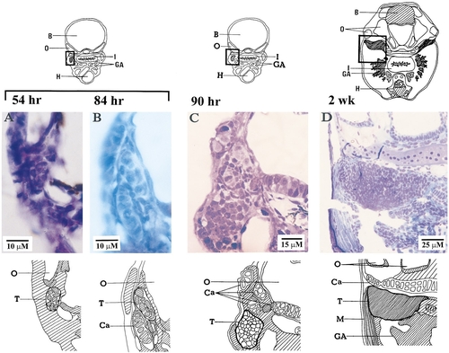
Histological development of zebrafish thymus. (A) Thymic primordium 54 hr after fertilization. Thymus at B, 84 hr; C, 90 hr; and D, 2 weeks. The diagram above each photograph shows its location and the sketch below identifies the organs. Photos A, B, and D are of paraffin sections stained with hematoxylin and eosin Y. Photo C is of a plastic section stained with toluidine blue. B, brain; Ca, cartilage; GA, gill arches; H, heart; I, intestine; M, muscle; O, otic vesicle; T, thymus. |

The thymus at 1 month. (A) The thymic epithilium is continuous with the pharyngeal epithilium (location of section is shown in diagram at left). (B) At higher magnification, thymocytes appear as groups of packed cells between nonlymphoid cells, including epithelial cells. Photos are of plastic sections stained with toluidine blue. B, brain; GA, gill arches; H, heart; I, intestine; O, otic vesicle; P, pharynx; T, thymus. Lymphoblasts, solid arrows marked ‘‘1’’; small lymphocytes, open arrows marked ‘‘2’’; epithelial cells, double arrowheads marked ‘‘3.’’ |
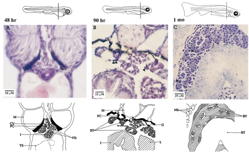
Histological development of zebrafish pronephros. (A) 48 hr, pronephric ducts associated with caudal vein. (B) 90 hr, renal tubules and glomerulae are visible; isolated cells resembling either primitive hematopoietic cells or lymphocyte-like cells appear (▲). (C) 1 month, hematopoietic tissue occupies the space between large renal tubules and blood vessels. The diagram above the photo shows the plane of each section and the sketch below identifies the organs. Photos A and B are of paraffin sections stained with hematoxylin and eosin Y; photo C is of a plastic section stained with toluidine blue. BV, blood vessel; CV, caudal vein; G, glomerlus; I, intestine; L, liver; M, muscle; Mb, muscle bundles; PC, pigment cell; PD, pronephric duct; RT, renal tubule; YS, yolk sac. |
Reprinted from Developmental Biology, 182(2), Willett, C.E., Zapata, A.G., Hopkins, N.A., and Steiner, L.A., Expression of zebrafish rag genes during early development identifies the thymus, 331-341, Copyright (1997) with permission from Elsevier. Full text @ Dev. Biol.