
|
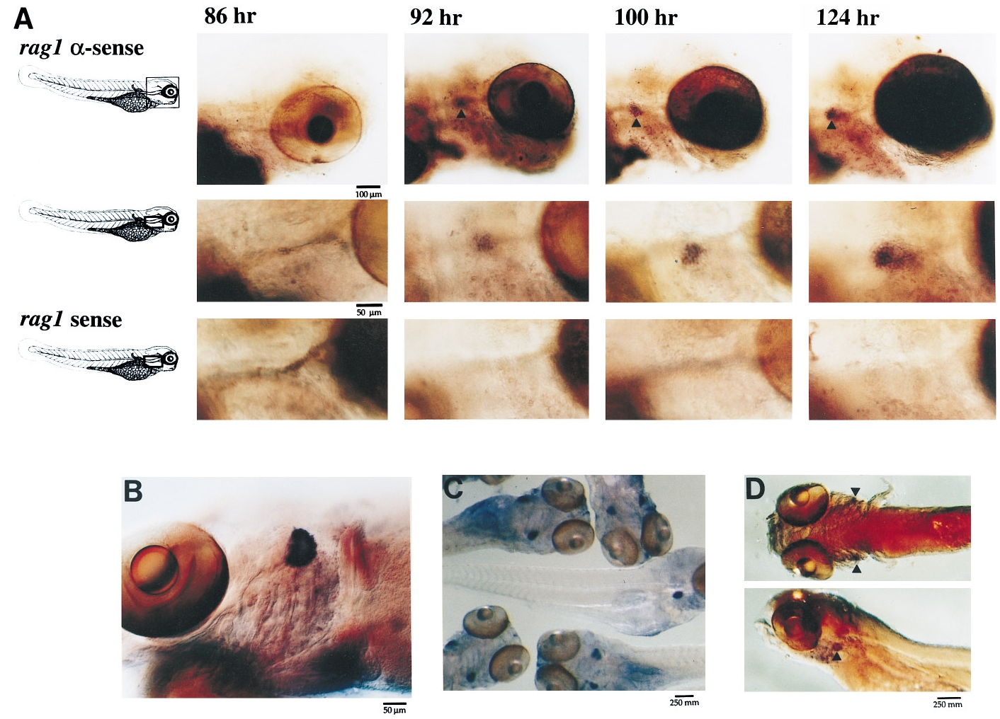
Fig. 1 Whole-mount in situ hybridization of rag expression during zebrafish development. (A) Embryos and larvae were harvested at 3- hr intervals and hybridized with either a rag1 sense or antisense probe. Staining with the rag1 antisense probe shows expression in bilaterally symmetric spherical regions posterior to the eye. Top row, original magnification 55x; middle row, original magnification 110x; bottom row, staining with the rag1 sense control (original magnification 110x). (B) rag1 whole-mount in situ hybridization of a 2- week-old fish. (C) rag1 staining of several 1-week-old fish. (D)A 7-day-old fish hybridized to rag2 under the same whole-mount hybridization conditions used for rag1.
Reprinted from Developmental Biology, 182(2), Willett, C.E., Zapata, A.G., Hopkins, N.A., and Steiner, L.A., Expression of zebrafish rag genes during early development identifies the thymus, 331-341, Copyright (1997) with permission from Elsevier. Full text @ Dev. Biol.