
FIGURE
Fig. 3
- ID
- ZDB-FIG-090511-17
- Publication
- Willett et al., 1997 - Expression of zebrafish rag genes during early development identifies the thymus
- Other Figures
- All Figure Page
- Back to All Figure Page
Fig. 3
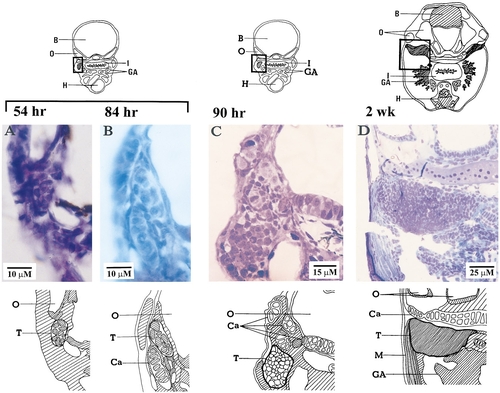
Histological development of zebrafish thymus. (A) Thymic primordium 54 hr after fertilization. Thymus at B, 84 hr; C, 90 hr; and D, 2 weeks. The diagram above each photograph shows its location and the sketch below identifies the organs. Photos A, B, and D are of paraffin sections stained with hematoxylin and eosin Y. Photo C is of a plastic section stained with toluidine blue. B, brain; Ca, cartilage; GA, gill arches; H, heart; I, intestine; M, muscle; O, otic vesicle; T, thymus. |
Expression Data
Expression Detail
Antibody Labeling
Phenotype Data
Phenotype Detail
Acknowledgments
This image is the copyrighted work of the attributed author or publisher, and
ZFIN has permission only to display this image to its users.
Additional permissions should be obtained from the applicable author or publisher of the image.
Reprinted from Developmental Biology, 182(2), Willett, C.E., Zapata, A.G., Hopkins, N.A., and Steiner, L.A., Expression of zebrafish rag genes during early development identifies the thymus, 331-341, Copyright (1997) with permission from Elsevier. Full text @ Dev. Biol.