
- Title
-
The Zpr-3 Antibody Recognizes the 320-354 Region of Rho and Labels Both Rods and Green Cones in Zebrafish
- Authors
- Hu, H., Qin, Y., Qu, Z., Huang, Y., Ren, X., Liu, M., Liu, F., Gao, P.
- Source
- Full text @ Zebrafish

The antigen of zpr-3 is zebrafish Rho protein. (A) The ladder-like bands produced by zpr-3 and the anti-Rho antibody in the western blot assays of wild-type zebrafish protein lysates. (B) Full-length zebrafish Rho protein tagged with Flag was expressed in ARPE-19 cells and detected by western blot using the zpr-3, anti-Rho and anti-Flag antibodies, respectively. The images of western blot are shown. (C) Overlapping of the fluorescence signals of zpr-3 and anti-Flag antibodies in ARPE-19 cells transfected with the zebrafish rho expression plasmid. Scale bar, 20 μm. |
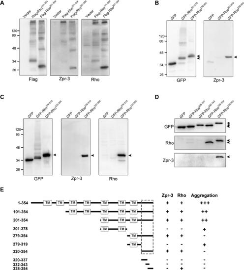
Identification of the exact epitope of zpr-3. Truncated zebrafish Rho proteins, 101–354 and 201–354 (A), 201–278 and 279–354 (B), 279–319 and 320–354 (C), 320–337, 332-343, and 338–354 (D), were expressed in ARPE-19 cells and detected by western blot using the zpr-3 and anti-Rho antibodies, respectively. The images of western blot are shown. Black arrows mark specific protein bands. (E) The schematic summary of the above western blot results is shown. The multiple transmembrane domains of Rho were indicated, and the epitope of zpr-3 was highlighted by the dashed frame |

The fluorescence signals of zpr-3 are not solely located in rods. (A) Immunofluorescence results of zpr-3 and the anti-Rho antibody in 14 dpf zebrafish retinal sections. Scale bar, 20 μm. (B) Partial co-localization of the red fluorescence signals of zpr-3 with the EGFP-labeled rods (arrows) in 7 dpf zebrafish retinal sections. Scale bar, 20 μm. (C) High-magnification views of the EGFP-labeled rod cells and the zpr-3-labeled outer segments of rods and other photoreceptors. Scale bar, 20 μm. EXPRESSION / LABELING:
|

Zpr-3 labels green cones as well as rods in immunofluorescence assays. (A) No primary antibody incubation was used as a negative control staining. (B) Double-label immunofluorescence images of zpr-3 and anti-Opn1lw antibody on zebrafish retinal sections. The fluorescence signals of zpr-3 were almost not co-localized with the outer segments of red-cones. Scale bar, 20 μm. N = 3 (biological replicates); n = 3 (technical replicates). (C) No primary antibody incubation was used as a negative control staining. (D) Double-label immunofluorescence images of zpr-3 and anti-Opn1mw antibodies on zebrafish retinal sections. Obvious co-localization between the fluorescence signals of zpr-3 and the outer segments of green cones is shown. Scale bar, 20 μm. N = 3 (biological replicates); n = 3 (technical replicates). INL, inner nuclear layer; NP, no primary antibody control; ONL, outer nuclear layer; OS, outer segment. (E) Phylogenetic tree of zpr-3 (320–354 aa in Rho) was reconstructed using the neighbor-joining method (left). Sequence alignment of the epitope of zpr-3 (320–354 aa in Rho) and its corresponding regions in eight opsins of cones (Opn1mw1-4, Opn1lw1-2, Opn1sw1, and Opn1sw2). The identical amino acid residues are marked in red (right). EXPRESSION / LABELING:
|