
Image
|
Figure Caption
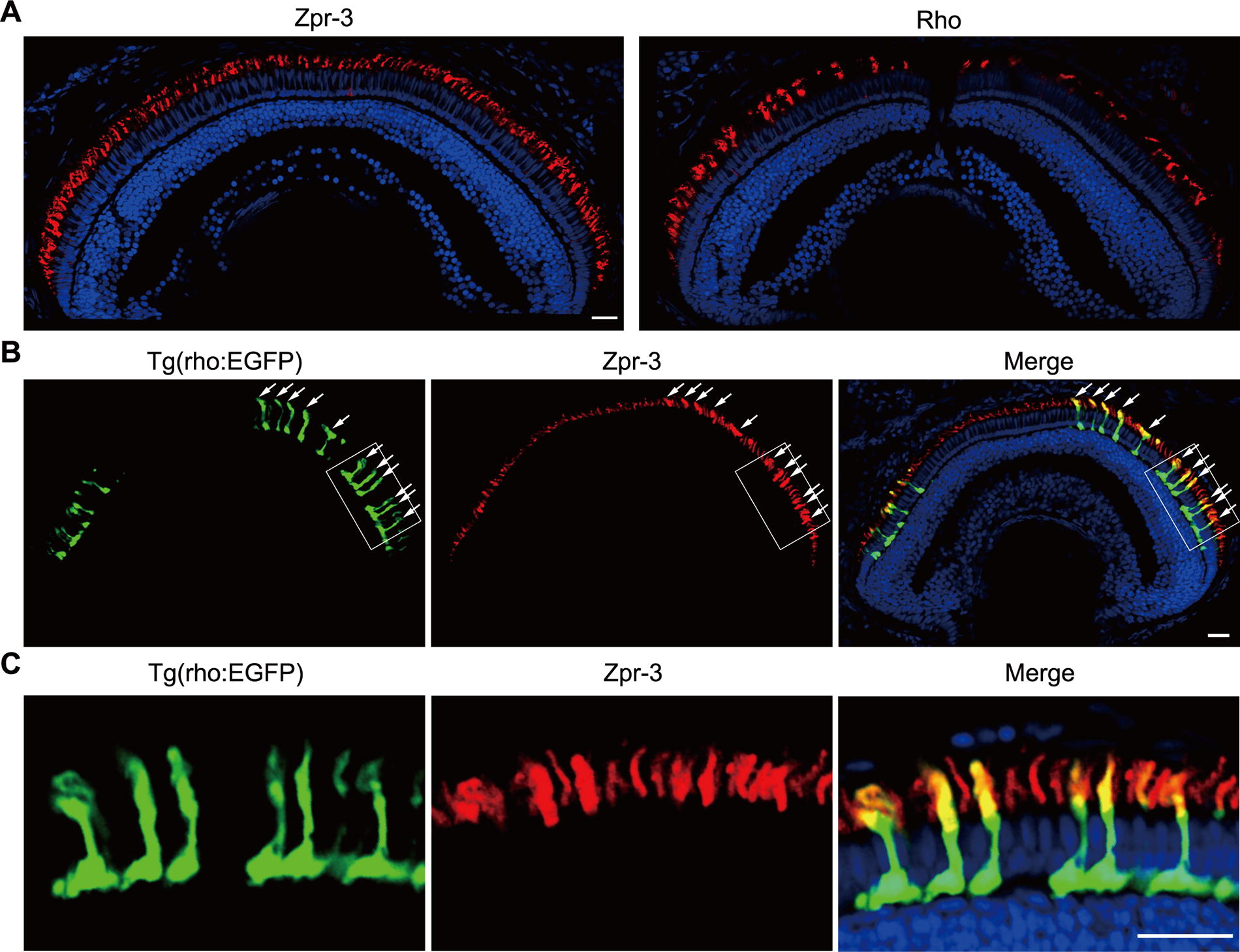
Fig. 3 The fluorescence signals of zpr-3 are not solely located in rods. (A) Immunofluorescence results of zpr-3 and the anti-Rho antibody in 14 dpf zebrafish retinal sections. Scale bar, 20 μm. (B) Partial co-localization of the red fluorescence signals of zpr-3 with the EGFP-labeled rods (arrows) in 7 dpf zebrafish retinal sections. Scale bar, 20 μm. (C) High-magnification views of the EGFP-labeled rod cells and the zpr-3-labeled outer segments of rods and other photoreceptors. Scale bar, 20 μm.
Figure Data
Acknowledgments
This image is the copyrighted work of the attributed author or publisher, and
ZFIN has permission only to display this image to its users.
Additional permissions should be obtained from the applicable author or publisher of the image.
Full text @ Zebrafish