FIGURE SUMMARY
Title

A zebrafish model of Ifih1-driven Aicardi-Goutières syndrome reproduces the interferon signature and the exacerbated inflammation of patients

Authors
Bernal-Bermúdez, B., Martínez-López, A., Martínez-Morcillo, F.J., Tyrkalska, S.D., Martínez-Menchón, T., Mesa-Del-Castillo, P., Cayuela, M.L., Mulero, V., García-Moreno, D.
Source
Full text @ Front Immunol

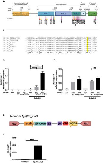
Localization, sequence alignments, ifih1 mRNA overexpression, and Tg(ifih1_mut) line design. (A) Protein diagram showing the positions of protein domains and their amino acid boundaries within the 1,025 residues of IFIH1 (MDA5) protein. The 28 previously published mutations are annotated. CARD, caspase activation recruitment domain; Hel, helicase domain, where Hel1 and Hel2 are the two conserved core helicase domains and Hel2i is an insertion domain that is conserved in the RIG-I–like helicase family; P, pincer or bridge region connecting Hel2 to the C-terminal domain (CTD) involved in binding double-stranded RNA. Red (24), yellow (2), purple (22), black (16), blue (4), and light purple (23). (B) Protein sequence alignment of IFIH1 from different species; IFIH1_ZEBRAFISH (ENSDART00000003913.10), IFIH1_CHICKEN (ENSGALT00010050768.1), IFIH1_MOUSE (ENSMUST00000028259.12), IFH1_PIG (ENSSSCT00000061806.3), IFIH1_HUMAN (ENST00000649979.2), IFH1_DOG (ENSCAFT00845053824.1), IFIH1_CAT (ENSFCAT00000026735.4). (C, D) RT-qPCR of isg15(C) or ifih1(D) from 3-dpf zebrafish larvae injected with wild-type (WT) or mutant (p.Arg742His) ifih1 mRNA with or without 25 pg/egg of poly I:C. (E) Scheme showing zebrafish p.Arg742His transgenic (Tg) design Tg(ifih1_mut) and (F) expression of ifih1 in the transgenic line Tg(ifih1_mut) by RT-PCR. p-values were calculated using one-way ANOVA and Tukey multiple range test. ns, not significant; ****p ≤ 0.0001.

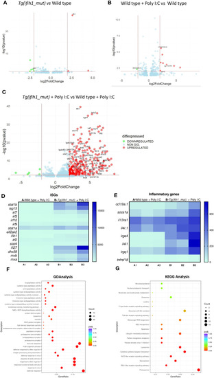
Transcriptomic analysis of the Tg(ifih1_mut) reveals systemic inflammation and an ISG signature upon poly I:C induction. Differentially expressed genes (DEGs) between wild-type larvae and Tg(ifih1_mut)(A), between wild type + Poly I:C and wild-type (B), and between Tg(ifih1_mut) + Poly I:C and wild type + Poly I:C (C). (D, E) Heatmap of analyzed ISGs (D) and proinflammatory genes (E) between Tg(ifih1_mut) + Poly I:C and wild type + Poly I:C represented by counts. (F, G) GO (F) and KEGG (G) analysis between Tg(ifih1_mut) + Poly I:C and wild type + Poly I:C.

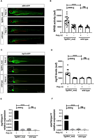
Real-time visualization of inflammation and type I IFN induction in Tg(ifih1_mut) upon poly I:C induction. nfkb:eGFP and isg15:eGFP (green heart marker) reporter zebrafish lines were crossed with Tg(ifih1_mut) (red eye marker) and injected or not with poly I:C. Nfkb activity (A, B) and isg15 levels (C, D) were analyzed by fluorescence microscopy and quantified. Each dot represents a larva and the mean ± SEM for each experimental group is also shown. Representative merge images of whole larvae were also shown (A, C). (E, F) Transcript levels of isg15 and stat1b in larvae from the cross of nfkb:eGFP and Tg(ifih1_mut) were analyzed by RT-qPCR and the results are shown as the mean ± SEM of pooled larvae. The region of interest (ROI) used to quantify the fluorescence in isg15:eGFP reporter line is indicated as a dot line in the images (C). p-values were calculated using one-way ANOVA and Tukey multiple range test. ns, not significant; ****p ≤ 0.0001.

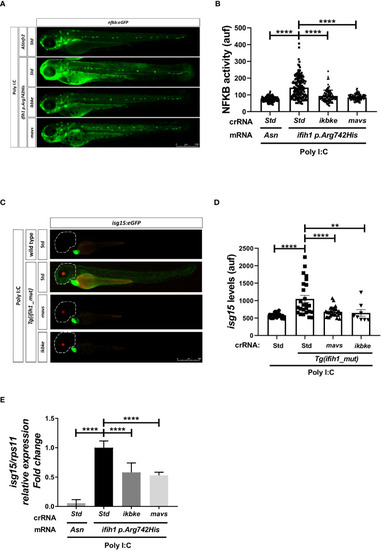
Genetic inhibition of Mavs/Ikbke signaling pathway impairs the induction of inflammation and ISGs in Tg(ifih1_mut) upon poly I:C stimulation. Tg(ifih1_mut) were crossed with nfkb:eGFP and isg15:eGFP reporter lines, injected with poly I:C and crRNA/Cas9 complexes for mavs or ikbke. Nfkb activity (A, B) and isg15 levels (C, D) were analyzed by fluorescence microscopy and quantified. Each dot represents a larva and the mean ± SEM for each experimental group is also shown. Representative merge images of whole larvae were also shown (A, C). (E) Transcript levels of isg15 in larvae from the cross of nfkb:eGFP and Tg(ifih1_mut) were analyzed by RT-qPCR and the results are shown as the mean ± SEM of pooled larvae. p-values were calculated using one-way ANOVA and Tukey multiple range test. **p ≤ 0.01, ****p ≤ 0.0001.

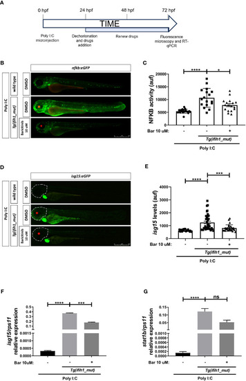
Pharmacological inhibition of Jak impairs the induction of ISGs and inflammation in
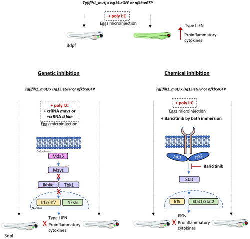
 Tg(ifih1_mut) upon poly I:C stimulation. (A)nfkb:eGFP and isg15:eGFP zebrafish were crossed with Tg(ifih1_mut), eggs were injected with poly I:C at 0 dpf, larvae were dechorionated at 1 dpf, and baricitinib was added to the water and renewed at 2 dpf, and images were taken and analyzed by microscopy at 3 dpf. (B–E) Nfkb activity (B, C) and isg15 levels (D, E) were analyzed by fluorescence microscopy and quantified. Each dot represents a larva and the mean ± SEM for each experimental group is also shown. Representative merge images of whole larvae were also shown (B, D). (F, G) Transcript levels of isg15(F) and stat1(G) in larvae from the cross of nfkb:eGFP and Tg(ifih1_mut) were analyzed by RT-qPCR and the results are shown as the mean ± SEM of pooled larvae. p-values were calculated using one-way ANOVA and Tukey multiple range test. ns, not significant; *p ≤ 0.05, **p ≤ 0.01, ***p ≤ 0.001, ****p ≤ 0.0001.

Schematic summary. Tg(ifih1_mut) crossed with the inflammation and type I interferon reporters, nfkb:eGFP and isg15:eGFP, activate the expression of GFP 3 days after the injection of poly I:C. Larvae bearing the mutation p.Arg742His show the red eye tag, and larvae bearing the isg15:eGFP reporter show the green heart tag marker. The genetic inhibition of mavs or ikbke prevents the transduction of the signal from Mda5 after sensing the poly I:C, avoiding the activation and translocation of Irf3/irf7 and Nfkb into the nucleus and preventing the expression of type I interferon and proinflammatory cytokines and the GFP activation. The chemical inhibition by the addition of Baricitinib by bath immersion provokes the blockade of the signal transduction through the Jak1 and Jak2 kinases, which results in the prevention of Stat and Irf9 translocation into the nucleus and activation of the ISGs and proinflammatory cytokine expression, avoiding the activation of GFP of the reporter lines.

Acknowledgments
This image is the copyrighted work of the attributed author or publisher, and ZFIN has permission only to display this image to its users. Additional permissions should be obtained from the applicable author or publisher of the image. Full text @ Front Immunol