FIGURE SUMMARY
Title

Evaluation of Neural Regulation and Microglial Responses to Brain Injury in Larval Zebrafish Exposed to Perfluorooctane Sulfonate

Authors
Paquette, S.E., Martin, N.R., Rodd, A., Manz, K.E., Allen, E., Camarillo, M., Weller, H.I., Pennell, K., Plavicki, J.S.
Source
Full text @ Environ. Health Perspect.

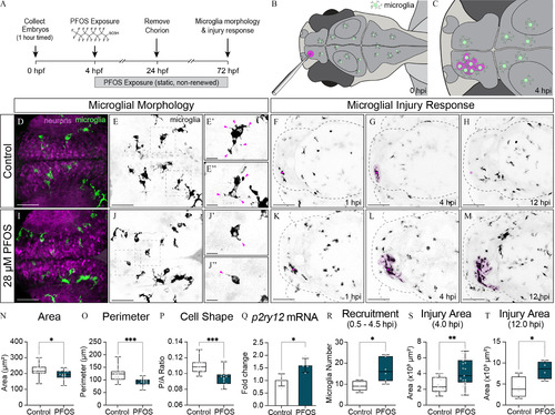
Microglia morphology and response to minor brain injury in 3-dpf larvae exposed to 28μM PFOS. (A) Exposure paradigm: zebrafish embryos from Tg(mpeg1:EGFP) adults were collected after a 1-h timed spawn. At 4 hpf, embryos were dosed with 0.1% DMSO (Control) or 28μM PFOS. Treatment solutions were static and not renewed. Embryos were dechorionated at 24 hpf, and imaging and injury experiments were conducted at 3 dpf. (B) Schematic of larval brain injury: a pulled-glass needle (OD 9μm) was used to puncture the right telencephalon of larval zebrafish. (C) Hypothetical schematic of microglia response 4 h post-injury (hpi). (D) Confocal micrographs of 3-dpf control-treated larvae with fluorescently labeled neurons and microglia. (E) Isolated fluorescently labeled microglia from (D). (E′–E″) 3× magnified microglia from boxes in (E). Magenta arrows point to projections emanating from the microglia cell bodies. (F) Dorsal view of a representative 3-dpf control-treated larval brain at 1 hpi. Injury site marked with magenta dot. (G) Dorsal view of a representative 3-dpf control-treated larval brain at 4 hpi. (H) Dorsal view of a representative 3-dpf control-treated larval brain at 12 hpi. (I) Confocal micrographs of 3-dpf 28μM PFOS-treated larvae with fluorescently labeled neurons and microglia. (J) Micrographs from (I) isolating just the fluorescently labeled microglia. (J′–J″) 3× magnified microglia from boxes in (J). Magenta arrows point to projections emanating from the microglia cell bodies. (K) Dorsal view of a representative 3-dpf 28μM PFOS-treated larval brain at 1 hpi. Injury site marked with magenta dot. (L) Dorsal view of a representative 3-dpf 28μM PFOS-treated larval brain at 4 hpi. (M) Dorsal view of a representative 3-dpf 28μM PFOS-treated larval brain at 12 hpi. (N) Quantification of microglia cell area. (O) Quantification of microglia cell perimeter. (P) Perimeter-to-area ratio of control-treated and PFOS-treated microglia. n=15 fish per group; 3–18 cells counted per fish. (Q) qRT-PCR results assessing the relative expression of the microglia activation gene, p2ry12, in isolated heads of control- and PFOS-treated 3-dpf larvae. n=4 samples of 10 pooled heads. (R) Number of microglia recruited to the injury site between 0.5 and 4.5 hpi. n=56 per group. (S) Quantification of the area of microglia response around the injury site at 4 hpi, as shown as the magenta overlays in (G) and (I). n=1920 fish per group. (T) Quantification of the area of microglia response around the injury site at 12 hpi. n=4 fish per group. Confocal micrographs at 40× magnification (D–E″ and I–J″) or 20× magnification (F–H; K–M). Error bars represent standard deviation. Box plot limits represent 25th to 75th percentile, with the midline representing the median. *p<0.05; **p<0.01; ***p<0.001. See Excel Table S1 for additional statistical details. Note: DMSO, dimethyl sulfoxide; dpf, days post fertilization; hpf, hours postfertilization; PFOS, perfluorooctane sulfonate.

Optogenetic modulation of PFOS-exposed microglia. (A) Schematic of halorhodopsin: Optogenetic modulation of microglia electrical state is achieved via photostimulation of the light-gated chloride pump, halorhodopsin (eNpHR3.0). eNpHR3.0 is most responsive to 589-nm wavelength light. (B) eNpHR3.0 was driven under a pan-macrophage promoter [Tg(mpeg1:GalFF;UAS:eNpHR3.0-mCherry)] to achieve optogenetic control of microglia in zebrafish larvae. (C) Experimental paradigm: At 72 hpf, injured or uninjured zebrafish were stimulated for 4 h with 589-nm light in the enclosed Noldus DanioVision Behavior Unit. (D) Confocal micrograph of 3-dpf zebrafish brain exposed to 28μM PFOS. Magenta cells are unstimulated halorhodopsin+ microglia. (E) Pseudocolored (D) in black and white. (E′–E″) 3× magnification of microglia from boxes in (E). Arrows point to projections emanating from the microglia cell bodies. (F) Dorsal view of a representative 3-dpf control-treated larval brain at 4 hpi without stimulation. Injury site marked with magenta dot and area of responding microglia shaded in magenta. (G) Dorsal view of a representative 3-dpf 28μM PFOS-treated larval brain at 4 hpi without stimulation. (H) Confocal micrograph of 3-dpf 28μM PFOS-treated larval brain following stimulation with 589-nm light. Magenta cells are stimulated halorhodopsin+ microglia. (I) Pseudocolored (H) in black and white. (I′–I″) 3× magnification of microglia from boxes in (I). Arrows point to projections emanating from the microglia cell bodies. (J) Dorsal view of a representative 3-dpf control-treated larval brain at 4 hpi following 589-nm light stimulation. (K) Dorsal view of a representative 3-dpf 28μM PFOS-treated larval brain at 4 hpi following 589-nm light stimulation. (L) Quantification of microglia cell area of 3-dpf 28μM PFOS-treated larvae with and without light stimulation. (M) Quantification of microglia cell perimeter of 3-dpf 28μM PFOS-treated larvae with and without light stimulation. (N) Perimeter-to-area ratio of individual microglia of 3-dpf 28μM PFOS-treated larvae with and without light stimulation. n=4042 cells per group from three independent experiments. (O) Quantification of the area of microglia response around the injury site at 4 hpi. n=915 fish per group. Confocal micrographs at 40× magnification (D–E″ and H–I″) or 20× magnification (F,G,J,K). *p<0.05; **p<0.01; ****p<0.0001. Error bars represent standard deviation. Box plot limits represent 25th to 75th percentile, with the midline representing the median. See Excel Table S1 for additional statistical details. Note: dpf, days post fertilization; hpf, hours postfertilization; ns, not significant; PFOS, perfluorooctane sulfonate.

Analysis of regional and global neuronal network activity following chronic exposure to PFOS. (A) Illustrative representation of a larval zebrafish brain with anatomical regions outlined: forebrain (FB), optic tectum (OT), cerebellum (Ce), hindbrain (HB), and whole brain (WB). (B) Illustrative representation of neuron-driven CaMPARI: neurons with low intracellular calcium (low Ca2+) remain green following 1-min exposure to 405-nm light, whereas neurons with high intracellular calcium (high Ca2+) photoconvert to red. (C) Confocal micrograph of a 3-dpf larvae expressing neuron-specific CaMPARI [Tg(elavl1:CaMPARI)] following 1 min photoconversion. (D) Generated high-intensity look-up-table (LUT) heat map of the red, photoconverted channel in (C) depicting high-activity neurons. Low-to-high intracellular calcium is depicted by a blue–red–white spectrum. Confocal micrographs of active neurons at 3-dpf in (E) control and (F) 7μM PFOS- and (G) 28μM PFOS-exposed larvae following 1-min photoconversion with 405-nm light. Neuronal activity can be quantified by determining the ratio of fluorescent intensity in the red vs. green channels (FRed/FGreen). (H) Quantification of the regional and global (i.e., WB) neuronal activity as normalized FRed/FGreen ratios of 3-dpf control or 7μM PFOS-treated larvae. (I) Quantification of the regional and global neuronal activity of 3-dpf control or 28μM PFOS-treated larvae. Confocal micrographs of active neurons at 5 dpf in (J) control and (K) 7μM PFOS- and (L) 14μM PFOS-exposed larvae following 1-min photoconversion with 405-nm light. (M) Quantification of the regional and global neuronal activity of 5-dpf control or 7μM PFOS-treated larvae. (I) Quantification of the regional and global neuronal activity of 5-dpf control or 14μM PFOS-treated larvae. Confocal micrographs at 10× magnification. n=2123 fish per group. **p<0.01; ***p<0.001. Error bars represent standard deviation. Box plot limits represent 25th to 75th percentile, with the midline representing the median. See Excel Table S1 for additional statistical details. Note: Ca2+, calcium ion; CaMPARI, calcium-modulated photoactivatable ratiometric integrator; dpf, days postfertilization; PFOS, perfluorooctane sulfonate.

Assessing anxiety-like swim behavior in PFOS-exposed larvae. (A) Larval swim behavior during a 30-min light/dark behavioral assay was used to determine time spent in the well’s center (crossing center: less anxious) vs. time spent along the well’s edge (center avoidance: more anxious). (B) Heat maps were generated to indicate mobile (blue) vs. stationary (red) swim activity within the well, as well as the zones traversed within the well. (C) Quantification of the time that 3-dpf 7μM, 14μM, or 28μM PFOS-treated larvae spent in the center of the well relative to control-treated larvae. Images are of representative heat maps from each treatment group. (D) Quantification of the time that 5-dpf 7μM or 14μM PFOS-treated larvae spent in the center of the well relative to control-treated larvae. Images are of representative heat maps from each treatment group. n=76101 fish per group. *p<0.05; **p<0.01; ****p<0.0001. Error bars represent SEM. See Excel Table S1 for additional statistical details. Note: dpf, days postfertilization; PFOS, perfluorooctane sulfonate; SEM, standard error of the mean.

Light/dark behavioral assay and neuronal activity in microglia mutants exposed to PFOS. Wild-type (WT) and microglia-deficient irf8st96/st96 larvae dosed with either control or 7μM PFOS were assessed for potential changes in swim behavior and neuronal activity. (A) Light/dark behavioral assay of 5-dpf WT or irf8/ mutant control or PFOS-exposed larvae. (B) Assessment of the average distance moved of 5-dpf larvae at minute 15, which is the start of the second light-off cycle, at minute 30, which is the end of the second light-off cycle, and the percentage change in distance moved from minute 15 to minute 30. (C) Quantification of neuronal CaMPARI activity in the forebrain of 5-dpf control-treated WT, PFOS-treated WT, control-treated mutant, and PFOS-treated mutant larvae. (D) Quantification of neuronal CaMPARI activity in the optic tectum. (E) Quantification of neuronal CaMPARI activity in the cerebellum. (F) Quantification of neuronal CaMPARI activity in the hindbrain. (G) Quantification of neuronal CaMPARI activity in the whole brain. n=1422 fish per group for behavior; n=1316 fish per group for neuroimaging. **p<0.01; ***p<0.001; ****p<0.0001. Error bars represent standard deviation. Box plot limits represent 25th to 75th percentile, with the midline representing the median. See Excel Table S1 for additional statistical details. Note: CaMPARI, calcium-modulated photoactivatable ratiometric integrator; Ce, cerebellum; dpf, days postfertilization; FB, forebrain; HB, hindbrain; OT, optic tectum; PFOS, perfluorooctane sulfonate; WB, whole brain.

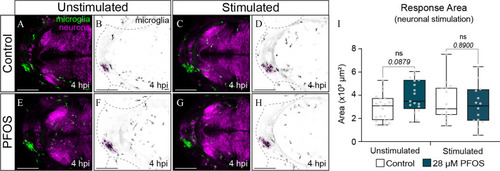
Microglia response to brain injury following optogenetic silencing of neurons in PFOS-exposed larvae. (A) Confocal micrograph of a 3-dpf control-treated larval brain with fluorescently labeled microglia in green and halorhodopsin+ neurons in magenta. Image taken 4 hpi without light stimulation. (B) Isolated microglia from (A), pseudocolored in black and white. Area of microglia response 4 hpi shaded with magenta. (C) Confocal micrograph of a 3-dpf control-treated larval brain with fluorescently labeled microglia in green and halorhodopsin+ neurons in magenta. Image taken 4 hpi following stimulation with 589-nm light. (D) Isolated microglia from (C), pseudocolored in black and white. (E) Confocal micrograph of a 3-dpf 28μM PFOS-treated larval brain with fluorescently labeled microglia in green and halorhodopsin+ neurons in magenta. Image taken 4 hpi without light stimulation. (F) Isolated microglia from (E), pseudocolored in black and white. (G) Confocal micrograph of a 3-dpf 28μM-treated larval brain with fluorescently labeled microglia in green and halorhodopsin+ neurons in magenta. Image taken 4 hpi following stimulation with 589-nm light. (H) Isolated microglia from (G), pseudocolored in black and white. (I) Quantification of the area of microglia response around the injury site at 4 hpi, as shown by the magenta overlays. n=1420 fish per group. Error bars represent standard deviation. Box plot limits represent 25th to 75th percentile, with the midline representing the median. See Excel Table S1 for additional statistical details. Note: dpf, days postfertilization; hpi, hours post-injury; ns, not significant; PFOS, perfluorooctane sulfonate.

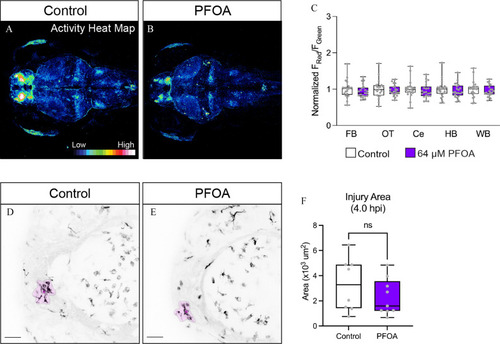
Neuronal activity and microglia response to brain injury following exposure to PFOA, a known immunotoxic congener. PFOA is an 8-carbon PFAS compound with a carboxylate head group. To observe the effects of PFOA on neuronal activity, we again used larvae of the Tg(elavl3:CaMPARI) background. (A) Representative heat map of neuronal activity of a 5-dpf control-treated larval brain. (B) Representative heat map of neuronal activity of a 5-dpf 64μM PFOA-treated larval brain. (C) Quantification of the regional and global (i.e., WB) neuronal activity as normalized FRed/FGreen ratios of 5-dpf control or 64μM PFOA-treated larvae. (D) Confocal micrograph of a 3-dpf control-treated larval brain with fluorescently labeled microglia. Image taken 4 hpi, with area of responding microglia shaded in magenta. (E) Confocal micrograph of a 3-dpf 64μM PFOA-treated larval brain with fluorescently labeled microglia. Image taken 4 hpi. (F) Quantification of the area of microglia response around the injury site at 4 hpi, as shown by the magenta overlays. n=89 fish per group. Error bars represent standard deviation. Box plot limits represent 25th to 75th percentile, with the midline representing the median. See Excel Table S1 for additional statistical details. Note: Ce, cerebellum; dpf, days postfertilization; FB, forebrain; HB, hindbrain; hpi, hours post-injury; ns, not significant; OT, optic tectum; PFAS, per- and polyfluoroalkyl substances; PFOA, perfluorooctanoic acid; WB, whole brain.

Acknowledgments
This image is the copyrighted work of the attributed author or publisher, and ZFIN has permission only to display this image to its users. Additional permissions should be obtained from the applicable author or publisher of the image. Full text @ Environ. Health Perspect.