FIGURE SUMMARY
Title

Functional analysis of germline VANGL2 variants using rescue assays of vangl2 knockout zebrafish

Authors
Derrick, C.J., Szenker-Ravi, E., Santos-Ledo, A., Alqahtani, A., Yusof, A., Eley, L., Coleman, A.H.L., Tohari, S., Ng, A.Y., Venkatesh, B., Alharby, E., Mansard, L., Bonnet-Dupeyron, M.N., Roux, A.F., Vaché, C., Roume, J., Bouvagnet, P., Almontashiri, N.A.M., Henderson, D.J., Reversade, B., Chaudhry, B.
Source
Full text @ Hum. Mol. Genet.

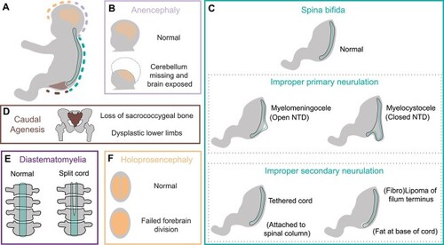
Congenital malformations associated with VANGL2 coding sequence missense mutations. (A) Regions associated with abnormal development in patients carrying heterozygous coding sequence mutations in VANGL2. (B) Anencephaly, failure of anterior neural tube closure during primary neurulation, leading to loss of cerebellum and open cranial cavity. (C) Different forms of spina bifida arising due to failure of primary neurulation which can result in an open or closed spinal column. VANGL2 patients also display failures in secondary neurulation: tethered cord, where free movement of the spinal cord within the column is restricted, and lipoma at the base of the spinal cord (filum terminus). (D) Caudal agenesis (also known as caudal regression syndrome or sacral agenesis) is defined by the loss of the sacrococcygeal bone (brown), associated with dysplastic lower limbs and impacts other caudally located organs. (E) Diastematomyelia is the splitting of the spinal cord. (F) Failure of division of the embryonic forebrain results in holoprosencephaly.

Collation of clinical data relating to historical VANGL2 variants. (A) Primary structure of human VANGL2 (UniProt: Q9ULK5) [116, 117] showing known functional domains and location of VUS. Two N-terminal serine/threonine motifs (cyan [78]), 4 transmembrane domains (magenta, [98]), TGN (trans-Golgi network) sorting motif (yellow [118]), the Dishevelled and Prickle binding region (green [119–121], which overlaps with two ubiquitinoylation sites, (orange [122], a VCP interacting motif (mauve, [122])) and a nuclear localization signal (white [123]). Two further ubiquitinoylation sites (orange) and the Type I PDZ domain (blue, [124]) are present at the C-terminal end. (B) Clinical pedigrees, major developmental defects and original references relating to previously reported missense mutations in coding sequence of human VANGL2. Squares, circles, diamonds, triangles represent male, females, unknown gender, and foetuses, respectively. Arrowheads denote proband. Diagonal line denotes deceased. Open symbols: healthy, black symbols: affected. V2: VANGL2 genotype, D2: DVL2 genotype. Question mark denotes DNA unavailable. Het.: heterozygous.

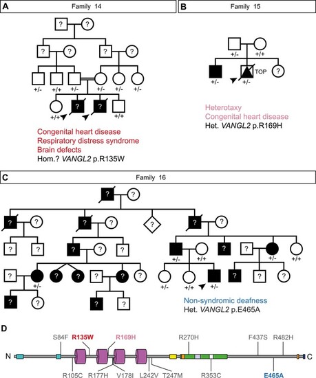
Identification of new VANGL2 germline variants and families. (A) Clinical pedigrees of putative recessive p.R135W VANGL2 family. Both deceased male probands (for which DNA was unavailable) possessed complete atrioventricular septal defect, respiratory distress syndrome, large occipital encephalocele and congenital hydrocephalus. Heterozygous family members were clinically normal. (B) Clinical pedigree of heterozygous p.R169H VANGL2 family. Antenatal diagnosis of male proband identified dextrocardia, persistent left superior vena cava, total anomalous pulmonary venous drainage, atrioventricular septal defect, transposition of great arteries, pulmonary atresia, right pulmonary isomerism and asplenia. The elder brother is also heterozygous with situs inversus. The heterozygous father was clinically normal. (C) Clinical pedigree of heterozygous p.E465A VANGL2 family in which proband has autosomal dominant non-syndromic hearing loss. The heterozygous p.E465A variant is also present in an unaffected relative. (D) Primary structure of Human VANGL2 showing known functional domains and location of previously reported and novel VUS reproduced from Fig. 2A. Squares, circles, diamonds, triangles represent male, females, unknown gender, and foetuses, respectively. Arrowheads denote proband. Diagonal line denotes deceased. Open symbols: healthy, black symbols: affected. Question mark denotes DNA unavailable. Het.: heterozygous, Hom: homozygous, TOP: termination of pregnancy.

vangl2m209 convergent extension defect can be rescued by most VANGL2 VUS. (A–C) Representative brightfield images of (A) uninjected sibling, (B) uninjected homozygous vangl2m209 mutant, (C) homozygous vangl2m209 mutant injected with WT VANGL2 mRNA. (D) Quantification of AP length at 26 hours post fertilization (hpf) to examine efficacy of WT VANGL2 to rescue vangl2m209 phenotype. Colour-coded regions defined by uninjected vangl2m209 mutant data range: within/no rescue is grey, above/rescue is green and below/worsens is magenta. WT VANGL2 mRNA injection partially rescues AP axis length. Each dot denotes a single embryo. (E) Homozygous vangl2m209 mutant injected with p.L242V VANGL2 mRNA, (F) homozygous vangl2m209 mutant injected with p.R135W VANGL2 mRNA, (G) homozygous vangl2m209 mutant injected with p.R169H VANGL2 mRNA, (H) homozygous vangl2m209 mutant injected with p.T247M VANGL2 mRNA, (I) homozygous vangl2m209 mutant injected with p.R177H VANGL2 mRNA, (J) homozygous vangl2m209 mutant injected with p.R482H VANGL2 mRNA. (K) Quantification of AP length at 26 hpf in homozygous vangl2m209 mutants injected with mRNA encoding different VANGL2 VUS. Variants are ranked and grouped based on result of statistical test when compared to uninjected homozygous vangl2m209 mutants and homozygous vangl2m209 mutants injected with WT VANGL2 mRNA (see Supplementary Table 2 for statistical tests). Each dot denotes a single embryo. Colour coded regions and dots denote comparison against uninjected homozygous vangl2m209 mutants from panel D. A-C, E-J: Lateral views, anterior left. D, K: Brown-Forsythe and Welch ANOVA with multiple comparisons, Mean ± SEM, ****: p < 0.0001. Scale bars: 0.2 mm.

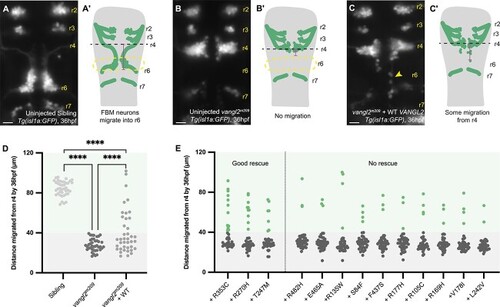
Motor neuron migration cannot be rescued by most
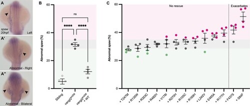
 VANGL2 variants. (A–A′) Representative hindbrain of uninjected Tg(isl1a:GFP) sibling at 36 hpf (A), motor neurons have migrated from rhombomere 4 (r4) into rhombomere 6 (r6). Measurement of migration distance (magenta line) from r4 (black line) schematized in (A′). (B–B′) Representative hindbrain of vangl2m209; Tg(isl1a:GFP) homozygous mutant at 36 hpf (B), motor neurons have failed to migrate from r4 into r6. Measurement of migration distance schematized in (B′). (C–C′) Representative hindbrain of vangl2m209; Tg(isl1a:GFP) homozygous mutant at 36 hpf injected with WT VANGL2 mRNA at 1-cell stage, some neurons exit r4 (arrowhead). Measurement of migration distance schematized in (C′). (D) Representation of Isl1a + neuron phenotype by migration distance from r4 at 36 hpf. Each dot denotes a single embryo. Colour-coded regions defined by uninjected homozygous vangl2m209 mutant data range: within/no rescue is grey, above/rescue is green. In homozygous vangl2m209 mutant embryos injected with WT VANGL2 mRNA, migration distance above the mutant range is defined as “rescue”. p-values represent Fisher’s exact test based on rescue and mutant categories for each group. WT VANGL2 mRNA injection partially rescues neuronal migration (see Supplementary Table 3 for statistical tests). (E) Representation of Isl1a + neuron phenotype by migration distance from r4 at 36 hpf in homozygous vangl2m209 mutants injected with mRNA encoding different VANGL2 VUS. Each dot denotes a single embryo, colour coded regions and dots denote comparison against the threshold of uninjected homozygous mutant data from panel D. Variants are ranked and grouped based on result of Fisher’s exact test when comparing rescue and mutant categories to uninjected homozygous vangl2m209 mutants and homozygous vangl2m209 mutants injected with WT VANGL2 mRNA (see Supplementary Table 4 for statistical tests). A–C: dorsal views, anterior up. D-E: Fisher’s exact tests, ****: p < 0.0001. Scale bars: 20 μm.

Asymmetric spaw expression in vangl2m209 mutants cannot be rescued by VANGL2 variants. (A–A″) Representative images of mRNA in situ hybridization for southpaw (spaw) the zebrafish homologue of Nodal at 20 hpf. In wild-type siblings (A), spaw is restricted to the left lateral plate mesoderm (LPM, arrowhead), alternatively, spaw may be abnormally expressed in the right LPM (A′, arrowhead) or bilaterally (A″, arrowheads). (B) Quantification of abnormal spaw expression at 20 hpf to examine efficacy of WT VANGL2 to rescue homozygous vangl2m209 mutant phenotype. WT VANGL2 mRNA injection completely rescues spaw expression. Colour-coded regions defined by vangl2m209 mutant data range: within/no rescue is grey, below/rescue is green and above/worsens is magenta. Each dot denotes a clutch, consisting of 17–30 embryos. (C) Quantification of abnormal spaw expression at 20 hpf in homozygous vangl2m209 mutants injected with mRNA encoding different VANGL2 VUS. Variants are ranked and grouped based on result of statistical test when compared to uninjected homozygous vangl2m209 mutants and homozygous vangl2m209 mutants injected with WT VANGL2 mRNA (see Supplementary Table 5 for statistical tests). Each dot denotes a clutch, consisting of 18–43 embryos, colour coded regions and dots denote comparison against the uninjected homozygous vangl2m209 mutant data from panel B. A–A″: Dorsal views, anterior up. B-C: One-way ANOVA with multiple comparisons, Mean ± SEM, ****: p < 0.0001.

Heart tube formation and positioning is sensitive to loss of
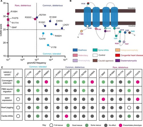
 vangl2 function. Representative heart tube phenotypes of homozygous vangl2m209 mutant at 26 hpf, highlighted by mRNA in situ hybridization of the pan cardiac marker myosin, light chain 7, regulatory (myl7). (A) Normal, left jogging (A′) right jogging (A″) no jogging and (A‴) cardia bifida, the presence of two shorter heart tubes at the midline. (B) Quantification of abnormal heart jogging scored live between 28–30 hpf to examine efficacy of WT VANGL2 to rescue homozygous vangl2m209 mutant phenotype. WT VANGL2 mRNA injection completely rescues heart jogging. Colour-coded regions defined by homozygous vangl2m209 mutant data range: within/no rescue is grey, below/rescue is green and above/worsens is magenta. Each dot denotes a clutch, consisting of 16–29 embryos. (C) Quantification of abnormal heart jogging scored live between 28–30 hpf in homozygous vangl2m209 mutants injected with mRNA encoding different VANGL2 VUS. Variants are ranked and grouped based on result of statistical test when compared to uninjected homozygous vangl2m209 mutants and homozygous vangl2m209 mutants injected with WT VANGL2 mRNA (see Supplementary Table 6 for statistical tests). Each dot denotes a clutch, consisting of 21–42 embryos, colour coded regions and dots denote comparison against the range of uninjected mutant data from panel B. (D) Quantification of cardia bifida scored live between 28–30hpf to examine efficacy of WT VANGL2 to rescue homozygous vangl2m209 phenotype. WT VANGL2 mRNA injection completely rescues cardia bifida. Colour-coded regions defined by homozygous vangl2m209 mutant data range: within/no rescue is grey, below/rescue is green and above/worsens is magenta. Each dot denotes a clutch, consisting of 16–29 embryos. (E) Quantification of cardia bifida scored live between 28–30 hpf in homozygous vangl2m209 mutants injected with mRNA encoding different VANGL2 VUS. Variants are ranked and grouped based on result of statistical test when compared to uninjected homozygous vangl2m209 mutants and homozygous vangl2m209 mutants injected with WT VANGL2 mRNA (see Supplementary Table 7 for statistical tests). Each dot denotes a clutch, consisting of 21–42 embryos, colour coded regions and dots denote comparison against uninjected homozygous vangl2m209 mutant data from panel D. Heart jogging and cardia bifida were analysed in the same clutch. A–A‴: dorsal views, anterior up. B-E: One-way ANOVA with multiple comparisons, Mean ± SEM, ****: p < 0.0001.

Summary of VANGL2 VUS in vivo activity. (A) Comparison of observed variant frequency and CADD score of VANGL2 variants. p.S84F, p.R169H, p.R177H and p.F437S have not been observed in the population and are assigned frequency of 10−7. (B) Schematic of VANGL2 protein and location of residues, amino acid (text) is colour coded based on (A), residue position is colour coded based on malformation associated with variant (Figs 2 and 3). (C) Summary table of vangl2-developmental processes that were able to be at least partially rescued by injection of WT VANGL2 mRNA and results of variant mRNA analysis. Full rescue denoted by open circles, good rescue: green, some rescue: dark green, no rescue: grey, and exacerbation: magenta.

Acknowledgments
This image is the copyrighted work of the attributed author or publisher, and ZFIN has permission only to display this image to its users. Additional permissions should be obtained from the applicable author or publisher of the image. Full text @ Hum. Mol. Genet.