
- Title
-
Znf598-mediated Rps10/eS10 ubiquitination contributes to the ribosome ubiquitination dynamics during zebrafish development
- Authors
- Ugajin, N., Imami, K., Takada, H., Ishihama, Y., Chiba, S., Mishima, Y.
- Source
- Full text @ RNA
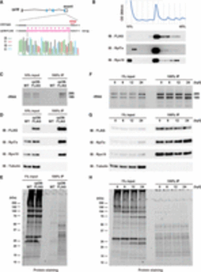
Establishment of a ribosome affinity purification system in zebrafish. (A) Scheme of a FLAG-tagged rpl36 gene locus. Genome sequences around the stop codon of wild-type (WT) and rpl36-FLAG strains are shown. The sequence chromatogram of rpl36-FLAG embryos is indicated below. (B) Representative polysome profiles of rpl36-FLAG embryos at 24 hpf. The distribution patterns of ribosomal proteins are indicated below. (C–E) Validation of the ribosome purification system with WT and rpl36-FLAG embryos at 24 hpf. Total lysates (input) and FLAG-immunoprecipitants (IPs) were subjected to RNA electrophoresis (C), immunoblotting analysis using antibodies against the indicated proteins (D), and protein staining (E). (F–H) Validation of the ribosome purification system during various developmental stages. The developmental time points are indicated above as hpf. Total lysates (input) and FLAG-IPs were subjected to RNA electrophoresis (F), immunoblotting analysis using antibodies against the indicated proteins (G), and protein staining (H). |
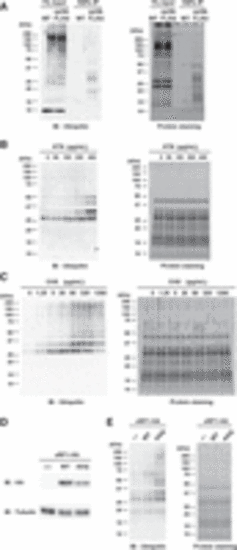
Detection of ribosome ubiquitination under normal and stress conditions. (A) Detection of ribosome ubiquitination. Total lysates (input) and FLAG-IPs obtained from WT and rpl36-FLAG embryos at 24 hpf were subjected to immunoblotting analysis with an anti-Ubiquitin antibody (left) and protein staining (right). (B) Analysis of ribosome ubiquitination under harringtonine (HTN) treatment. FLAG-IPs were subjected to immunoblotting analysis with an anti-Ubiquitin antibody (left) and protein staining (right). HTN concentrations are indicated above. (C) Analysis of ribosome ubiquitination under cycloheximide (CHX) treatment. FLAG-IPs were subjected to immunoblotting analysis with an anti-Ubiquitin antibody (left) and protein staining (right). CHX concentrations are indicated above. (D,E) Analysis of ribosome ubiquitination in the presence of mutant release factor eRF1-AAQ. Total lysates were subjected to immunoblotting analysis of eRF1-HA and Tubulin (D). FLAG-IPs were subjected to immunoblotting analysis with an anti-Ubiquitin antibody (E, left) and protein staining (E, right). |
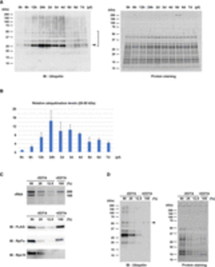
Ribosome ubiquitination level changes during zebrafish development. (A) Detection of ribosome ubiquitination during development. FLAG-IPs from various developmental stages were subjected to immunoblotting analysis with an anti-Ubiquitin antibody (left) and protein staining (right). Arrowhead indicates the most noticeable ubiquitination signal. Ubiquitination signals between 25 and 50 kDa were reproducibly detected (bracket). The developmental time points are indicated above as hpf or dpf. (B) A bar graph shows ubiquitination levels relative to 0 hpf. Ubiquitination signals between 25 and 50 kDa in (A, left) were normalized by corresponding protein amounts in (A, right). The average of three independent experiments is indicated. The error bars indicate the standard deviation. (C) Validation of 60S subunits purification. FLAG-IPs in the presence (+) or absence (−) of EDTA were subjected to RNA electrophoresis (upper) and immunoblotting analysis of ribosomal proteins (lower). The −EDTA samples were serially diluted as indicated above. (D) Detection of ribosome ubiquitination in the presence (+) or absence (−) of EDTA. FLAG-IPs in (C) were subjected to immunoblotting analysis with an anti-Ubiquitin antibody (left) and protein staining (right). Arrowhead indicates a ubiquitination signal derived from 60S subunits. |
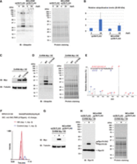
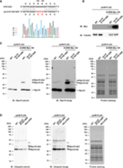
Znf598 promotes ribosome ubiquitination during development. (A) Comparison of ribosome ubiquitination levels of rpl36-FLAG and MZznf598;rpl36-FLAG embryos. FLAG-IPs from 0 and 24 hpf embryos were subjected to immunoblotting analysis with an anti-Ubiquitin antibody (left) and protein staining (right). The developmental time points are indicated above as hpf. (B) A bar graph shows ubiquitination levels relative to that of rpl36-FLAG embryos at 0 hpf. Ubiquitination signals between 25 and 50 kDa in (A, left) were normalized by corresponding protein amounts in (A, right). The average of three independent experiments is indicated. The error bars indicate the standard deviation. (C,D) Comparison of ribosome ubiquitination levels with or without Znf598 overexpression. Total lysates were subjected to immunoblotting analysis of Znf598-Myc and Tubulin (C). FLAG-IPs were subjected to immunoblotting analysis with an anti-Ubiquitin antibody (D, left) and protein staining (D, right). (E) A representative MS/MS spectrum of Rps10/eS10 di-glycyl K139. (F) MS-based quantification of a ubiquitinated peptide of Rps10/eS10 containing di-glycyl K139 under Znf598 OE (red) and control (blue) conditions. (G,H) Detection of Rps10/eS10 ubiquitination. Total lysates were subjected to immunoblotting analysis of Znf598-Myc and Tubulin (G). FLAG-IPs were subjected to immunoblotting analysis with an anti-Rps10 antibody (H, left) and protein staining (H, right). White and black arrowheads indicate nonubiquitinated or ubiquitinated Rps10/eS10 signals, respectively. The asterisk indicates a nonspecific signal. |
During zebrafish development, Rps10/eS10 ubiquitination is crucial for establishing the ribosome ubiquitination pattern. (A) Alignment of WT and K139/140R rps10 sequences. The sequencing chromatogram of the rps10 K139/140R embryos is indicated below. (B,C) Detection of Rps10/eS10 ubiquitination with or without Znf598 overexpression in rpl36-FLAG and rps10 K139/140R;rpl36-FLAG embryos. Total lysates were subjected to immunoblotting analysis of Znf598-Myc and Tubulin (B). FLAG-IPs were subjected to immunoblotting analysis with an anti-Rps10 antibody (C, left and middle) and protein staining (C, right). White and black arrowheads indicate nonubiquitinated or ubiquitinated Rps10/eS10 signals, respectively. Asterisks indicate nonspecific signals. (D) Comparison of ribosome ubiquitination pattern in rpl36-FLAG, rps10 K139/140R;rpl36-FLAG, and MZznf598;rpl36-FLAG embryos. FLAG-IPs were subjected to immunoblotting analysis with an anti-Ubiquitin antibody (left and middle) and protein staining (right). Black arrowheads indicate putative ubiquitinated Rps10/eS10 signals. White arrowheads indicate reduced ubiquitination signals in rps10 K139/140R;rpl36-FLAG and MZznf598;rpl36-FLAG embryos. |
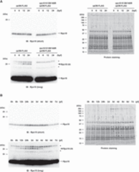
Rps10/eS10 ubiquitination level temporally changes during development. (A) Detection of Rps10/eS10 ubiquitination from 0 to 24 hpf embryos. FLAG-IPs were subjected to immunoblotting analysis with an anti-Rps10 antibody (left) and protein staining (right). The developmental time points are indicated above as hpf. White and black arrowheads indicate nonubiquitinated or ubiquitinated Rps10/eS10 signals, respectively. Asterisks indicate nonspecific signals. (B) Detection of Rps10/eS10 ubiquitination during development. FLAG-IPs were subjected to immunoblotting analysis with an anti-Rps10 antibody (left) and protein staining (right). The developmental time points are indicated above as hpf or dpf. White and black arrowheads indicate nonubiquitinated or ubiquitinated Rps10/eS10 signals, respectively. Asterisks indicate nonspecific signals. |