
Fig. 3
- ID
- ZDB-FIG-240201-58
- Publication
- Ugajin et al., 2023 - Znf598-mediated Rps10/eS10 ubiquitination contributes to the ribosome ubiquitination dynamics during zebrafish development
- Other Figures
- All Figure Page
- Back to All Figure Page
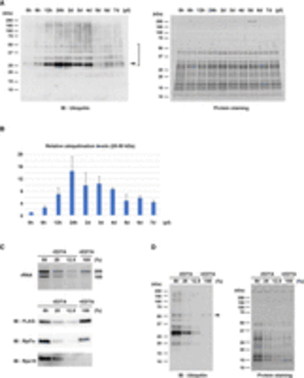
Ribosome ubiquitination level changes during zebrafish development. (A) Detection of ribosome ubiquitination during development. FLAG-IPs from various developmental stages were subjected to immunoblotting analysis with an anti-Ubiquitin antibody (left) and protein staining (right). Arrowhead indicates the most noticeable ubiquitination signal. Ubiquitination signals between 25 and 50 kDa were reproducibly detected (bracket). The developmental time points are indicated above as hpf or dpf. (B) A bar graph shows ubiquitination levels relative to 0 hpf. Ubiquitination signals between 25 and 50 kDa in (A, left) were normalized by corresponding protein amounts in (A, right). The average of three independent experiments is indicated. The error bars indicate the standard deviation. (C) Validation of 60S subunits purification. FLAG-IPs in the presence (+) or absence (−) of EDTA were subjected to RNA electrophoresis (upper) and immunoblotting analysis of ribosomal proteins (lower). The −EDTA samples were serially diluted as indicated above. (D) Detection of ribosome ubiquitination in the presence (+) or absence (−) of EDTA. FLAG-IPs in (C) were subjected to immunoblotting analysis with an anti-Ubiquitin antibody (left) and protein staining (right). Arrowhead indicates a ubiquitination signal derived from 60S subunits. |