
|
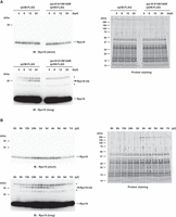
Fig. 6 Rps10/eS10 ubiquitination level temporally changes during development. (A) Detection of Rps10/eS10 ubiquitination from 0 to 24 hpf embryos. FLAG-IPs were subjected to immunoblotting analysis with an anti-Rps10 antibody (left) and protein staining (right). The developmental time points are indicated above as hpf. White and black arrowheads indicate nonubiquitinated or ubiquitinated Rps10/eS10 signals, respectively. Asterisks indicate nonspecific signals. (B) Detection of Rps10/eS10 ubiquitination during development. FLAG-IPs were subjected to immunoblotting analysis with an anti-Rps10 antibody (left) and protein staining (right). The developmental time points are indicated above as hpf or dpf. White and black arrowheads indicate nonubiquitinated or ubiquitinated Rps10/eS10 signals, respectively. Asterisks indicate nonspecific signals.