FIGURE SUMMARY
Title

Aicardi Syndrome Is a Genetically Heterogeneous Disorder

Authors
Ha, T.T., Burgess, R., Newman, M., Moey, C., Mandelstam, S.A., Gardner, A.E., Ivancevic, A.M., Pham, D., Kumar, R., Smith, N., Patel, C., Malone, S., Ryan, M.M., Calvert, S., van Eyk, C.L., Lardelli, M., Berkovic, S.F., Leventer, R.J., Richards, L.J., Scheffer, I.E., Gecz, J., Corbett, M.A.
Source
Full text @ Genes (Basel)

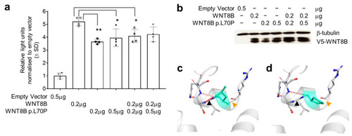
WNT8B p.Leu70Pro (p.L70P in this figure) affects protein function and structure. (a) TOPflash dual-luciferase reporter gene activity in HEK293T cells transfected with pcDNA3.1+ (empty vector) or pcDNA3.1+ expressing cDNAs for wildtype WNT8B, the p.Leu70Pro variant or combinations of both as shown below the horizontal axis. Data in the bars represent the mean and standard deviation of the ratio of firefly to Renilla luciferase luminescence normalised to the mean luminescence ratio in cells transfected with the empty vector from two independent transfections, each with technical duplicates per group. Individual replicate data are shown with point symbols for each bar. Comparisons were made using one-way ANOVA between all groups except the empty vector with significant differences when assessed using Tukey’s post hoc tests observed as shown * p < 0.05, ** p < 0.01. (b) Western blot showing the abundance of WNT8B wildtype and p.Leu70Pro variant proteins detected using rabbit anti-V5 antibody 500 ng/mL (Bethyl Cat# A190-120P, Montgomery, TX) in each transfection relative to β-tubulin detected using rabbit anti-β-tubulin antibody directly conjugated to HRP 200 ng/mL (Abcam Cat# ab21058, Cambridge, United Kingdom). (c) WNT8B compared to the predicted changes (d) in structure due to the p.Leu70Pro variant as assessed using DynaMut. Note that within the vicinity of the p.Leu70Pro variant there is loss of hydrogen bonding (black arrows) and loss of non-polar interactions (orange arrows).

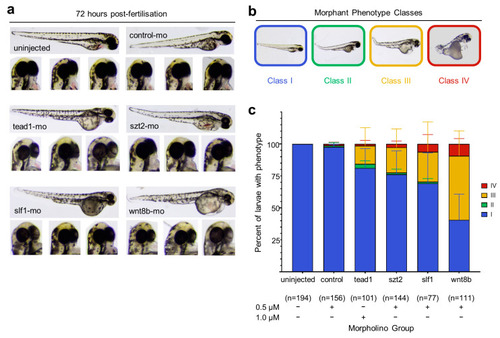
Phenotype analysis of morpholino mediated knockdown of Aicardi candidate genes in zebrafish embryos. (a) Examples of morphology of zebrafish embryos at 72 h post fertilization (hpf) either uninjected or injected with a control- or gene-targeted morpholino. Note the reduction in eye pigment in embryos injected with the tead1, slf1 or wnt8b morpholinos. (b) Zebrafish embryos were scored according to the classification system of Miesfeld et al. (2015) [58]. Class I: normal phenotype; Class II: normal eye pigmentation with mild body defects; Class III: reduced eye pigmentation with normal to mild body defects; Class IV; reduced eye pigmentation and severe body defects. (c) Observed morphant phenotypes after researcher-blinded injections of 240 zebrafish embryos per group. The number of viable embryos remaining after 72 hpf are denoted by (n). The amount of morpholino injected per group are indicated by (+).

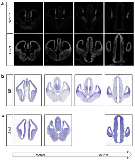
Wnt8b and Slf1 have spatially restricted expression patterns in the developing mouse brain. Coronal 20 μm frozen sections of mouse brain harvested at embryonic day 12. (a) Top row shows expression of Wnt8b measured using indirect immunofluorescent staining. Detection by rabbit anti-WNT8B primary antibody. Nuclei in the same section visualised using DAPI staining are shown in the second row to define morphology of the section. (b) Detection of Slf1 transcripts by in situ hybridisation using an in vitro transcribed anti-sense riboprobe. (c) Detection of Szt2 transcripts by in situ hybridisation using an in vitro transcribed anti-sense riboprobe.

Acknowledgments
This image is the copyrighted work of the attributed author or publisher, and ZFIN has permission only to display this image to its users. Additional permissions should be obtained from the applicable author or publisher of the image. Full text @ Genes (Basel)