IMAGE

Figure 1

ID
ZDB-IMAGE-230828-37
Source
Figures for Ha et al., 2023
Image
Figure Caption

Figure 1

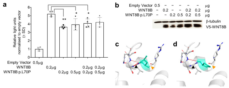
WNT8B p.Leu70Pro (p.L70P in this figure) affects protein function and structure. (a) TOPflash dual-luciferase reporter gene activity in HEK293T cells transfected with pcDNA3.1+ (empty vector) or pcDNA3.1+ expressing cDNAs for wildtype WNT8B, the p.Leu70Pro variant or combinations of both as shown below the horizontal axis. Data in the bars represent the mean and standard deviation of the ratio of firefly to Renilla luciferase luminescence normalised to the mean luminescence ratio in cells transfected with the empty vector from two independent transfections, each with technical duplicates per group. Individual replicate data are shown with point symbols for each bar. Comparisons were made using one-way ANOVA between all groups except the empty vector with significant differences when assessed using Tukey’s post hoc tests observed as shown * p < 0.05, ** p < 0.01. (b) Western blot showing the abundance of WNT8B wildtype and p.Leu70Pro variant proteins detected using rabbit anti-V5 antibody 500 ng/mL (Bethyl Cat# A190-120P, Montgomery, TX) in each transfection relative to β-tubulin detected using rabbit anti-β-tubulin antibody directly conjugated to HRP 200 ng/mL (Abcam Cat# ab21058, Cambridge, United Kingdom). (c) WNT8B compared to the predicted changes (d) in structure due to the p.Leu70Pro variant as assessed using DynaMut. Note that within the vicinity of the p.Leu70Pro variant there is loss of hydrogen bonding (black arrows) and loss of non-polar interactions (orange arrows).

Acknowledgments
This image is the copyrighted work of the attributed author or publisher, and ZFIN has permission only to display this image to its users. Additional permissions should be obtained from the applicable author or publisher of the image. Full text @ Genes (Basel)