
- Title
-
Stepwise modulation of apical orientational cell adhesions for vertebrate neurulation
- Authors
- Zhang, L., Wei, X.
- Source
- Full text @ Biol. Rev. Camb. Philos. Soc.

Orientational intercellular relationships and orientational cell adhesions (OCAs) in zebrafish neurulation. (A) Diagrams summarising characteristic changes in four aspects during the neural keel–rod–tube transition in zebrafish neurulation: orientational intercellular relationships between ipsilateral or contralateral cells (distinguished by pink and blue); spatiotemporal localization order of pioneer, intermediate, and terminal apical polarity proteins (represented by blue, red, and green dots, respectively; the blue dots in the neural keel represent only ZO-1 distribution because the pioneer proteins N-cadherins and E-cadherin are distributed on the entire lateral cell membranes at this stage); the morphological changes in apical surface alignments; and the dynamics of the apical OCAs. Black circles represent the contours of the neural keel, early and late neural rod, and neural tube. The grey boxes indicate the local regions that are magnified and presented in the diagrams below the neural tissue contours. Large black arrows indicate transition of developmental stages during neurulation; large and long blue, red, and green arrows indicate the progression and span of time when pioneer, intermediate, and terminal proteins are expressed, respectively. Small arrows indicate antiparallel or parallel apical OCAs (red trans-cell lines). Arrowheads indicate opposing apical OCAs (black trans-cell lines). aPKC, atypical protein kinase C; Crb, Crumbs; Lin7c, lineage abnormal 7c; Nok, Nagie oko; Pard3, partitioning defective 3; Pard6γb, partitioning defective 6 gamma b; ZO-1, zonula occludens-1. (B) Table showing the developmental and compositional changes of the OCAs. Parallel OCAs, adhesions at the lateral cell membranes between ipsilateral cells; antiparallel OCAs, adhesions at the lateral cell membranes between contralateral cells; parallel apical OCAs, four types of adhesions (enclosed by the bracket sign ‘[’) at the apical end of the lateral cell membranes between ipsilateral cells; opposing apical OCAs, adhesions between the apices of contralateral cells; AJ, adherens junction; Crb*, Crb-based adhesion; Na+/K+ ATPase*, Na+/K+-ATPase-based adhesion; TJ, tight junction; +, presence; −, absence. Modified from Guo et al. (2018). |

Parallel and antiparallel intercellular relationships at the lateral cell membranes of the neural cells change dynamically during neurulation. (A) Interdigitation of the apical protrusions of contralateral neural keel cells (pink and blue) generates the antiparallel orientational relationship between contralateral cells at the interdigitation zone, which is centred at the midline. This region may sense tension of cells. The balancing point of this tension (the spring icon) may automatically define the midline, thus acting as a midline sensor. The basal halves of neural keel cells display a parallel orientational relationship between ipsilateral cells. Corresponding antiparallel orientational cell adhesion (OCAs) and parallel OCAs are expected to exist in these regions that maintain the antiparallel and parallel intercellular relationships, respectively. (B) Plot illustrating that the parallel and antiparallel surface areas will change in opposite directions during the neural plate–keel–rod transition, with 1 representing the total lateral surface area. |
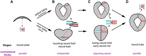
The conservation of cellular configurations and the orientational cell adhesions (OCAs) between contralateral cells in neural tube closure. (A) At the neural plate stage, all cells are oriented with their apical ends facing dorsally. This configuration implies that only parallel OCAs exist between neighbouring contralateral cells that flank the midline. This cellular configuration and OCAs are similar among all vertebrates. (B) Gradual folding and sharp folding change the neural plate to the neural fold in mammals (top) and the neural keel in teleosts (bottom). The two folding modes imply that the conserved antiparallel intercellular configuration exists either narrowly in the dorsal region of the neural fold or broadly in the neural keel. (C) The antiparallel intercellular relationship recedes and gives rise to an opposing apical intercellular relationship, with contralateral cells coalesced at the apices through opposing apical OCAs. Opposing apical OCAs require N-cadherin in zebrafish. Whether opposing apical OCAs exist in mammals is yet to be determined. (D) At the neural tube stage, neighbouring contralateral cells have coalesced at the dorsal roof and ventral floor by only parallel OCAs. |