
|
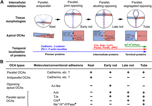
Fig. 1 Orientational intercellular relationships and orientational cell adhesions (OCAs) in zebrafish neurulation. (A) Diagrams summarising characteristic changes in four aspects during the neural keel–rod–tube transition in zebrafish neurulation: orientational intercellular relationships between ipsilateral or contralateral cells (distinguished by pink and blue); spatiotemporal localization order of pioneer, intermediate, and terminal apical polarity proteins (represented by blue, red, and green dots, respectively; the blue dots in the neural keel represent only ZO-1 distribution because the pioneer proteins N-cadherins and E-cadherin are distributed on the entire lateral cell membranes at this stage); the morphological changes in apical surface alignments; and the dynamics of the apical OCAs. Black circles represent the contours of the neural keel, early and late neural rod, and neural tube. The grey boxes indicate the local regions that are magnified and presented in the diagrams below the neural tissue contours. Large black arrows indicate transition of developmental stages during neurulation; large and long blue, red, and green arrows indicate the progression and span of time when pioneer, intermediate, and terminal proteins are expressed, respectively. Small arrows indicate antiparallel or parallel apical OCAs (red trans-cell lines). Arrowheads indicate opposing apical OCAs (black trans-cell lines). aPKC, atypical protein kinase C; Crb, Crumbs; Lin7c, lineage abnormal 7c; Nok, Nagie oko; Pard3, partitioning defective 3; Pard6γb, partitioning defective 6 gamma b; ZO-1, zonula occludens-1. (B) Table showing the developmental and compositional changes of the OCAs. Parallel OCAs, adhesions at the lateral cell membranes between ipsilateral cells; antiparallel OCAs, adhesions at the lateral cell membranes between contralateral cells; parallel apical OCAs, four types of adhesions (enclosed by the bracket sign ‘[’) at the apical end of the lateral cell membranes between ipsilateral cells; opposing apical OCAs, adhesions between the apices of contralateral cells; AJ, adherens junction; Crb*, Crb-based adhesion; Na+/K+ ATPase*, Na+/K+-ATPase-based adhesion; TJ, tight junction; +, presence; −, absence. Modified from Guo et al. (2018).