FIGURE SUMMARY
Title

Chronic Vitamin E Deficiency Dysregulates Purine, Phospholipid, and Amino Acid Metabolism in Aging Zebrafish Skeletal Muscle

Authors
Henderson, T.D., Choi, J., Leonard, S.W., Head, B., Tanguay, R.L., Barton, C.L., Traber, M.G.
Source
Full text @ Antioxidants (Basel)

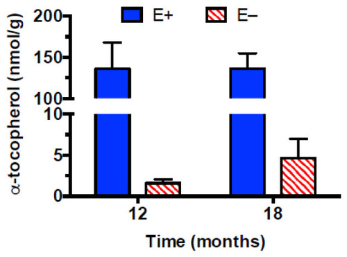
Muscle α-tocopherol concentrations. Zebrafish from the same cohort were fed a vitamin E-sufficient (E+, blue bars, n = 5 per group) or -deficient (E−, red striped bars, n = 5 at 12 months and n = 4 at 18 months) diet from age 45 days until 12 or 18 months. At the indicated times, fish were sacrificed, the muscles harvested, frozen, and kept frozen until analysis. α-Tocopherol skeletal muscle concentrations (mean ± SD) show a diet main effect (p ≤ 0.0001, no significant interaction or main effect of time).

Analysis of E+ fish muscles at 12 vs. 18 months. The heatmap (A) shows the top 25 metabolites separating groups and their relative abundances per sample (red represents a higher concentration per sample while blue indicates a lower one). The partial least squares-discriminant analysis (PLS-DA) scores plot (B) separates 12- and 18-month E+ muscle samples by feature differences and displays a 95% confidence region around samples. Important features plot displays variable importance in projection (VIP) (C) of the PLS-DA and provides an estimation of the discriminatory power of each metabolite with a score. Larger VIP scores indicate greater discriminatory power in the PLS-DA models. The table (D) lists the top values shown in the graph of pathway analysis, while figure (E) displays pathway analysis by ranking metabolites by p-value (shown as −log10(p)) and pathway impact referenced against the KEGG (Kiyoto Encyclopedia of Genes and Genomes) Pathway Database. Red indicates highest impacts, clear the lowest, yellow and orange are intermediate.

Metabolomic differences between E+ and E− fish muscles at 12 months. Shown are the heatmap (A) and PLS-DA scores plot (B), which separates E+ and E− muscle samples by feature differences and displays a 95% confidence region around samples and VIPs (C). There were insufficient statistically significant differences in the data from muscles of E+ and E− fish at 12 months for statistically meaningful pathway analysis.

Metabolomic differences between E+ and E− fish muscles at 18 months. Shown are the heatmap (A) PLS-DA scores plot (B) and VIP scores (C). The table (D) lists the top values shown in the graph of pathway analysis. (E) displays pathway analysis by ranking metabolites by p-value (shown as −log10(p)) and pathway impact referenced against the KEGG Pathway Database. Red indicates highest impacts, clear the lowest, yellow and orange are intermediate.

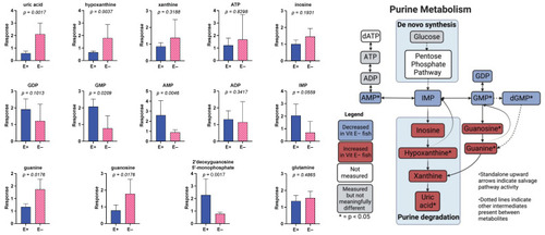
Purine metabolism is altered in E− zebrafish muscle at 18 months. Bar graphs showing Student’s t-test of 14 metabolites involved in KEGG pathway of zebrafish purine metabolism. Relative response (mean ± SD, n = 5 per group, E+ (blue bar), E− (red stripped bar), group = 18 months). The right graphic is a visualization of the changes in various purines within the purine metabolism pathway. Right graphic created with BioRender.com.

Metabolomic differences between E− fish at 12 and 18 months. Shown are the heatmap (A) PLS-DA scores plot (B,C) VIP scores. There was insufficient statistically significant differences in the data from muscles of E− fish at 12 and 18 months for statistically meaningful pathway analysis.

Acknowledgments
This image is the copyrighted work of the attributed author or publisher, and ZFIN has permission only to display this image to its users. Additional permissions should be obtained from the applicable author or publisher of the image. Full text @ Antioxidants (Basel)