
- Title
-
Loss of CSF-contacting neuron sensory function is associated with a hyper-kyphosis of the spine reminiscent of Scheuermann's disease
- Authors
- Marie-Hardy, L., Slimani, L., Messa, G., El Bourakkadi, Z., Prigent, A., Sayetta, C., Koëth, F., Pascal-Moussellard, H., Wyart, C., Cantaut-Belarif, Y.
- Source
- Full text @ Sci. Rep.
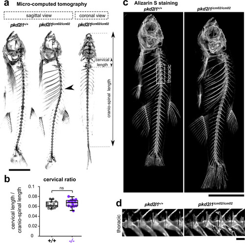
pkd2l1 null mutants develop during adulthood an idiopathic hyper-kyphosis of the thoracic spine. (a) 3D micro-computed tomography reconstruction images of a 18 months-old wild-type (pkd2l1+/+, left sagittal view) and a pkd2l1icm02/icm02 mutant sibling (middle, sagittal view; right, frontal view). pkd2l1icm2/icm02 mutants exhibit a thoracic hyper-kyphosis of the spine (arrowhead) but show no deformity in the coronal plane. Scale bar: 0.5 cm. (b) Distribution at 18 months-old of the cervical ratio, defined as the cervical length divided by the cranio-spinal length, in wild-type (grey, + / + , n = 13) and pkd2l1icm02/icm02 mutants (purple, −/−, n = 13). Each point represents a single animal. Boxplots represent median values ± IQR. ns: p > 0.05, Mann–Whitney test. (c) Sagittal views of Alizarin S stained-skeletal preparations of 18 months-old wild-type (pkd2l1+/+, left) and a pkd2l1icm02/icm02 mutant sibling (right). Scale bar: 0.5 cm. (d) High-magnification fluorescence images of Alizarin S in the thoracic region of the spine (as defined in (c), dotted line rectangle) in a wild-type (left) and a pkd2l1icm02/icm02 sibling (right). Neural arches (empty arrowhead), hemal arches (double empty arrowhead) and ribs (solid arrowhead) are highlighted for the pkd2l1icm02/icm02 mutant. Note that in both genotypes, no vertebral dislocation, malformation or fusion are observed. Scale bar: 500 µm. PHENOTYPE:
|
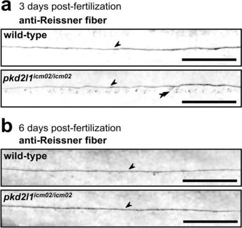
pkd2l1 mutant larvae retain a normally assembled Reissner fiber in the central canal of the spinal cord. Sagittal views of representative immunostainings against the Reissner fiber material imaged in the central canal of the spinal cord of 3 dpf (a) and 6 dpf (b) wild-type (top, one representative larva out of 8/9 at 3/6 dpf, respectively), and pkd2l1icm02/icm02 mutants (bottom, one representative larva out of 7/9 at 3/6 dpf, respectively). Arrows denote the presence of a continuous Reissner fiber in the central canal of the spinal cord. Double arrowheads indicate the presence of Reissner fiber-positive material in the floor plate at 3 dpf. Larvae are oriented rostral to the left and dorsal to the top. Scale bars: 50 µm. PHENOTYPE:
|
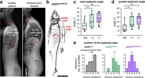
Pkd2l1 loss-of-function induces a hyper-kyphosis of the thoracic spine reminiscent of Scheuermann’s disease. (a) X-ray radiographs in the sagittal plane of a healthy patient (left; male, 20 years old) and of a patient with Scheuermann’s idiopathic disease (right; male, 20 years old). The deformity of the thoracic spine (right) is characterized by a Cobb angle between the first (T1) and the twelfth (T12) thoracic vertebra of 70° and is associated with no vertebral malformations. (b) Representative 3D micro-computed tomography reconstructions of a pkd2l1icm02/icm02 mutant (18 months-old) used to exemplify the evaluation in the sagittal plane of the main kyphosis angle, the greater kyphosis angle and the kyphosis apex. Scale bar: 0.5 cm. (c, d) Distribution of the main kyphosis angle (c) and greater kyphosis angle (d) in wild-type (black, + / + , n = 13), heterozygous (green, + /−, n = 10) and pkd2l1icm02/icm02 mutants (purple, −/−, n = 13). Boxplots represent median values ± IQR. Each point represents a single animal. *: p < 0.05, **: p < 0.01, Mann–Whitney test. (e) Distribution of the position of the apex of the kyphotic curve. The average position of the kyphosis apex is represented for each genotype (arrow, mean ± SEM). PHENOTYPE:
|
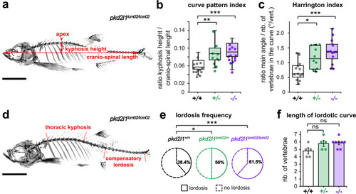
Adult pkd2l1 mutants develop a sagittal imbalance of the spine. (a) Representative 3D micro-computed tomography reconstruction of a pkd2l1icm02/icm02 mutant (18 months-old) used to exemplify the evaluation in the sagittal plane of the kyphosis height, the cranio-spinal length and the kyphosis apex. Scale bar 0.5 cm. (b, c) Distribution of the curve pattern index (b) and Harrington index (c) in wild-type (black, + / + , n = 13), heterozygous (green, + /−, n = 10) and pkd2l1icm02/icm02 mutants (purple, −/−, n = 13). Boxplots represent median values ± IQR. Each point represents a single animal. *: p < 0.05, **: p < 0.01, ***: p < 0.001, Mann–Whitney test. (d) Representative 3D micro-computed tomography reconstruction used to identify in the sagittal plane the thoracic kyphosis and a compensatory lordosis in the caudal region of the spine, exemplified in a 18 months-old pkd2l1icm02/icm02 mutant. Scale bar: 0.5 cm. (e) Pie charts representing the frequency of caudal lordotic curves in wild-type (black, n = 13), heterozygous (green, n = 10) and pkd2l1icm02/icm02 mutants (purple, n = 13). *: p < 0.05, ***: p < 0.001, Chi-Square test. (f) Histogram representing the length of the compensatory curve in lordotic wild-type (grey, + / + , n = 5), heterozygous (green, + /−, n = 5) and pkd2l1icm02/icm02 animals (purple, −/−, n = 6). Each point represents a single animal. Histograms represent mean values ± SEM. ns: p > 0.05, Mann–Whitney test. PHENOTYPE:
|
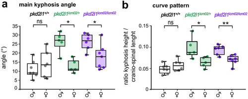
The severity of spine hyper-kyphosis in pkd2l1 mutants is sexually biased towards males. Distribution of the main kyphosis angle (a) and the curve pattern (b) in 18 months-old males and females of wild-type (black, + / + , n = 7 and 6 males and females respectively), heterozygous (green, + /, n = 5 and 5 males and females respectively) and mutant genotypes (purple, −/−, n = 6 and 7 males and females respectively). Each point represents a single animal. Boxplots represent median values ± IQR. *: p < 0.05, **: p < 0.01, Mann–Whitney test. PHENOTYPE:
|