
Figure 2
- ID
- ZDB-IMAGE-230406-17
- Publication
- Marie-Hardy et al., 2023 - Loss of CSF-contacting neuron sensory function is associated with a hyper-kyphosis of the spine reminiscent of Scheuermann's disease
- All Figures
- Figures for Marie-Hardy et al., 2023
|
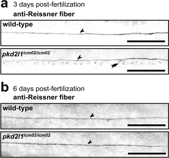
Figure 2
pkd2l1 mutant larvae retain a normally assembled Reissner fiber in the central canal of the spinal cord. Sagittal views of representative immunostainings against the Reissner fiber material imaged in the central canal of the spinal cord of 3 dpf (a) and 6 dpf (b) wild-type (top, one representative larva out of 8/9 at 3/6 dpf, respectively), and pkd2l1icm02/icm02 mutants (bottom, one representative larva out of 7/9 at 3/6 dpf, respectively). Arrows denote the presence of a continuous Reissner fiber in the central canal of the spinal cord. Double arrowheads indicate the presence of Reissner fiber-positive material in the floor plate at 3 dpf. Larvae are oriented rostral to the left and dorsal to the top. Scale bars: 50 µm.