FIGURE SUMMARY
Title

Therapeutic targeting of vascular malformation in a zebrafish model of hereditary haemorrhagic telangiectasia

Authors
Snodgrass, R.O., Govindpani, K., Plant, K., Kugler, E.C., Doh, C., Dawson, T., McCormack, L.E., Arthur, H.M., Chico, T.J.A.
Source
Full text @ Dis. Model. Mech.

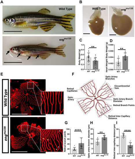
Adult engmu130 fish display cutaneous and retinal vascular malformations similar to those in patients with hereditary haemorrhagic telangiectasia and an enlarged heart. (A) Adult 5- to 6-month-old engmu130 zebrafish display cutaneous vascular malformations (black arrows). Scale bars: 1 cm. (B) Representative engmu130 and wild-type (WT) hearts (n=5/group). Scale bars: 500 μm. (C) Body weight of adult WT and engmu130 zebrafish. (D) Cardiac ventricle weight/body weight of WT and engmu130 zebrafish. (E) Fluorescent micrographs of whole-mount retinae from Tg(kdrl:Hsa.HRAS-mCherry)s916 zebrafish, showing abnormal vessel communications in engmu130 (white arrowheads). Scale bars: 500 μm. (F) Schematic diagram of a 5- to 6-month-old zebrafish retinal vasculature. Figure created with BioRender. (G) Number of retinal vascular branches in WT and engmu130 homozygous siblings. (H) Optic artery diameter in WT and engmu130 homozygous siblings. (I) Inter-capillary distance in WT and engmu130 homozygous siblings. **P<0.01, ****P<0.0001 (unpaired two-tailed Student's t-test, 8-10 animals/group).

engmu130 mutant embryos have increased basilar artery diameter and increased number of endothelial kugeln. (A) Representative maximum-intensity projection of Tg(kdrl:Hsa.HRAS-mCherry)s916 zebrafish brain vasculature at 72 hpf. Black arrowheads indicate individual kugeln. Scale bars: 150 μm/50 μm (inset). Three-dimensional reconstruction of the same image shown in green is on the right. (B) Representative maximum-intensity projection of the basilar artery (BA) at 72 hpf. Black arrowheads indicate individual kugeln. White bars indicate where diameter of the BA was measured. Scale bar: 50 μm. Three-dimensional reconstructions of the same images shown in green are on the right. (C) BA diameter in engmu130 mutants and WT siblings (unpaired two-tailed Student's t-test, 10-12 animals/group). (D) eng mutation did not alter endothelial cell (EC) number in the BA (unpaired two-tailed Student's t-test, 10-12 animals/group). (E) Kugel number per animal was increased in engmu130 mutants at 52 and 74 h post fertilisation (hpf) but not at 100 hpf. *P<0.05; ns, not significant (Mann–Whitney U-test, 8-27 animals/group).

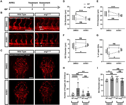
Vegf inhibition between 2 and 3 dpf rescues the abnormal trunk and cerebral vessel phenotypes of engmu130 mutants. (A) Experimental plan for rescuing the eng mutant phenotype in zebrafish using AV951. dpf, days post fertilisation. (B) Representative maximum-intensity projection of the trunk vasculature of engmu130 and WT embryos±AV951 (25 nM) treatment for 24 h. DA, dorsal aorta; PCV, posterior cardinal vein. Scale bar: 150 μm. (C) Representative maximum-intensity projection of the cerebral vasculature of engmu130 and WT embryos±AV951 (25 nM) treatment for 24 h. Scale bar: 100 μm. (D) DA diameter in engmu130 and WT embryos±AV951 treatment. (E) PCV diameter in engmu130 and WT embryos±AV951 treatment. (F) Intersegmental blood vessel (ISV) diameters in engmu130 and WT embryos±AV951 treatment. (G) Percentage of open ISVs in engmu130 and WT embryos±AV951 treatment. (H) Number of kugeln in engmu130 and WT embryos±AV951 treatment. (I) BA diameter in engmu130 and WT embryos±25 nM AV951 treatment. *P<0.05, **P<0.01, ***P<0.001, ****P<0.0001; ns, not significant (two-way ANOVA with Tukey post-hoc test, 10 animals/group).

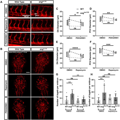
MEK or TOR inhibition normalises the vascular phenotype of engmu130 embryos. WT and engmu130 mutant embryos were treated with the MEK inhibitor PD0325901 (10 µM) or with the TOR inhibitor rapamycin (2.5 µM) for 24 h between 2 and 3 dpf, or with DMSO vehicle. (A) Representative maximum-intensity projections of the trunk vessels of engmu130 and WT embryos±PD0325901 (10 µM) or rapamycin (2.5 µM) treatment for 24 h. Scale bar: 150 μm. (B) Representative maximum-intensity projections of the cerebral vessels of engmu130 and WT embryos±PD0325901 (10 µM) or rapamycin (2.5 µM) treatment for 24 h. Scale bar: 100 μm. (C,D) engmu130 mutant embryos show reduced DA (C) and PCV (D) diameter following PD0325901 treatment, with PCV diameter normalised to WT values. (E,F) engmu130 mutant embryos show reduced DA diameter (E) and PCV diameter (F) following rapamycin treatment, with PCV diameter normalised to WT values. (G) engmu130 mutant embryos show normalised kugel formation following PD0325901 treatment. (H) engmu130 mutant embryos show normalised kugel formation following rapamycin treatment. *P<0.05, **P<0.01, ***P<0.001, ****P<0.0001; ns, not significant (two-way ANOVA with Tukey post-hoc test, 10-14 animals/group).

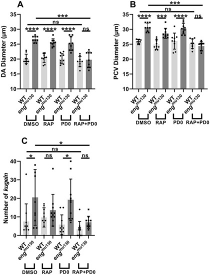
Combined low-dose mTOR and MEK inhibition prevents abnormal trunk vasculature and excess kugel formation in engmu130 zebrafish embryos. (A-C) Neither 2 µM rapamycin (RAP) nor 7.5 µM PD0325901 (PD0) alters the vascular phenotype of engmu130 mutants, but combined treatment resulted in normalised DA and PCV diameters, as well as reduced kugel formation in engmu130 zebrafish embryos. *P<0.05, ***P<0.001, ****P<0.0001; ns, not significant (two-way ANOVA with Tukey post-hoc test, 10-14 animals/group).

Acknowledgments
This image is the copyrighted work of the attributed author or publisher, and ZFIN has permission only to display this image to its users. Additional permissions should be obtained from the applicable author or publisher of the image. Full text @ Dis. Model. Mech.