IMAGE

Fig. 4.

ID
ZDB-IMAGE-230420-81
Genes
Source
Figures for Snodgrass et al., 2023
Image
Figure Caption

Fig. 4.

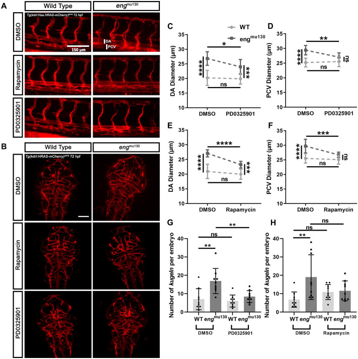
MEK or TOR inhibition normalises the vascular phenotype of engmu130 embryos. WT and engmu130 mutant embryos were treated with the MEK inhibitor PD0325901 (10 µM) or with the TOR inhibitor rapamycin (2.5 µM) for 24 h between 2 and 3 dpf, or with DMSO vehicle. (A) Representative maximum-intensity projections of the trunk vessels of engmu130 and WT embryos±PD0325901 (10 µM) or rapamycin (2.5 µM) treatment for 24 h. Scale bar: 150 μm. (B) Representative maximum-intensity projections of the cerebral vessels of engmu130 and WT embryos±PD0325901 (10 µM) or rapamycin (2.5 µM) treatment for 24 h. Scale bar: 100 μm. (C,D) engmu130 mutant embryos show reduced DA (C) and PCV (D) diameter following PD0325901 treatment, with PCV diameter normalised to WT values. (E,F) engmu130 mutant embryos show reduced DA diameter (E) and PCV diameter (F) following rapamycin treatment, with PCV diameter normalised to WT values. (G) engmu130 mutant embryos show normalised kugel formation following PD0325901 treatment. (H) engmu130 mutant embryos show normalised kugel formation following rapamycin treatment. *P<0.05, **P<0.01, ***P<0.001, ****P<0.0001; ns, not significant (two-way ANOVA with Tukey post-hoc test, 10-14 animals/group).

Figure Data
Acknowledgments
This image is the copyrighted work of the attributed author or publisher, and ZFIN has permission only to display this image to its users. Additional permissions should be obtained from the applicable author or publisher of the image. Full text @ Dis. Model. Mech.