
- Title
-
Distinct and redundant roles for zebrafish her genes during mineralization and craniofacial patterning
- Authors
- Stenzel, A., Mumme-Monheit, A., Sucharov, J., Walker, M., Mitchell, J.M., Appel, B., Nichols, J.T.
- Source
- Full text @ Front Endocrinol (Lausanne)

her6 and her9 are expressed in the same subpopulation of cranial neural crest cells as jag1b, and their expression is dependent upon jag1b function. (A) UMAPs from single cell RNA-sequencing on 24 hpf sorted cranial neural crest cells. As in our previous work (29), cranial neural crest cells can be clustered into four distinct populations. Based on our previous study and the landmark genes enriched in each population we can identify where the cells reside in the intact animal. Arrows indicate jag1b enrichment in a subset of anterior arch cells. Although her6 is broader, arrows indicate that some of the strongest expressing cells are the jag1b enriched populations. her9 is also broadly expressed among cranial neural crest cells, but at lower levels than her6. Arrows indicate her9 enrichment in the jag1b;her6 expressing populations. (B) We performed qPCR on 28 and 48 hpf genotyped heads from wild type and jag1b homozygous mutant siblings. Expression of each her gene was normalized to rps18, then normalized to the relevant wild-type control. Reactions were performed in technical triplicate and the results represent three to six biological replicates. Each biological replicate was pooled heads from 5–6 genotyped homozygous wild types or homozygous mutants. Error bars are standard deviation. Asterisks indicate significant differences from wild type controls at p < 0.05 by students t-test. |
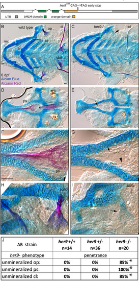
her9 is required for efficient mineralization. (A) Exon diagram for her9 indicating the untranslated regions (UTR) and the regions predicted to encode the basic helix-loop-helix (bHLH) and Orange domains. The location of the early stop encoded by the her9co66 allele is indicated. (B, C) Zebrafish heterozygous for her9 were pairwise intercrossed and six days post fertilization (dpf) larvae were stained with Alcian Blue and Alizarin Red to label cartilage and bone. The individuals were then genotyped, the viscerocrania were dissected, flat mounted, and then imaged. The following craniofacial skeletal elements are indicated in the wild-type individual: opercle bone (op), branchiostegal ray (br), maxilla (mx), entopterygoid (en), ceratobranchial 5 (cb5) bone, teeth (t), and the cleithrum (cl). Arrow in C indicates the unmineralized, Alcian Blue-positive opercle. Scale bar is 50 μm (D, E) The neurocrania from the same experiments as B and C were dissected, flat mounted, and then imaged. The parasphenoid bone (ps) and notochord (n) is indicated in the wild-type individual. Arrow in D indicates the shadow bone where the mineralized parasphenoid would be observable in wild types. (F, G) Enlargements from D and E. Arrow indicates the unmineralized shadow bone. Arrowhead denotes osteoblast-shaped cells surrounding the shadow bone. Scale bar is 50 μm (H, I) Enlargements from (B, C). The adductor operculi and the levetor operculi muscles (ao/lo) are indicated. Arrow in I indicates the unmineralized, Alcian Blue-positive opercle with the adductor operculi and the levetor operculi muscles (ao/lo) attached. (J) The penetrance of her9 mutant-associated phenotypes observed in 6 dpf larvae for each genotype are indicated. Asterisk indicates significant difference (p<0.05) in penetrance between the indicated genotype and wild type by Fishers exact test. PHENOTYPE:
|
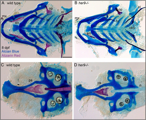
Mineralization defects in her9 homozygous mutants partially recover at late stages of larval development. Zebrafish heterozygous for her9 were pairwise intercrossed and at eight days post fertilization (dpf) larvae were stained with Alcian Blue and Alizarin Red to label cartilage and bone. The individuals were then genotyped, the viscerocrania and neurocrania were dissected, flat mounted, and then imaged. (A, B) Viscerocrania from wild-type and her9 homozygous mutant larvae. (C, D) Neurocrania from wild-type and her9 homozygous mutant larvae. The following craniofacial elements are indicated: opercle bone (op), branchiostegal ray (br), maxilla (mx), entopterygoid (en), ceratobranchial 5 (cb5) bone, teeth (t), cleithrum (cl), parasphenoid bone (ps), and notochord (n). Scale bar is 100 μm. PHENOTYPE:
|
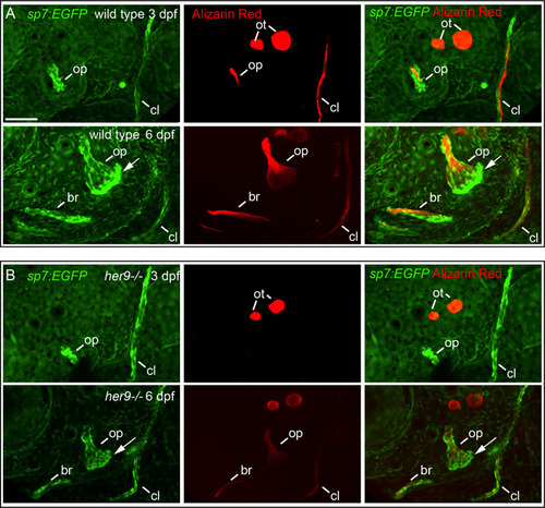
Osteoblasts differentiate in a spatiotemporal pattern similar to wild types in her9 mutants, but only weakly mineralize bone. sp7:EGFP;her9 heterozygotes were crossed to her9 heterozygotes and offspring were sorted for transgene expression. 16 live transgenic animals were labeled with Alizarin Red and imaged at 3 dpf. After imaging, animals were recovered and grown in individual wells until 6 dpf when they were labeled with Alizarin Red and imaged again. Individual animals were recovered for genotyping to identify homozygous wild types (A) and homozygous her9 mutants (B) that were imaged twice. The following structures are indicated: opercle bone (op), branchiostegal ray (br), cleithrum (cl), otoliths (ot). Arrows indicate osteoblasts prominently detected along the ventral edge of the opercle at 6 dpf. Scale bar is 50 μm. PHENOTYPE:
|
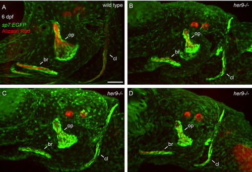
Opercle shapes are variable in her9 mutants. sp7:EGFP;her9 heterozygotes were crossed to her9 heterozygotes and offspring were sorted for transgene expression. 24 live transgenic animals were labeled with Alizarin Red and imaged at 6 dpf. Imaged animals were genotyped. (A) Representative opercle region from a wild-type larva. (B–D) Three different her9 mutant larvae are shown. The following structures are indicated: opercle bone (op), branchiostegal ray (br), cleithrum (cl). Scale bar is 50 μm. PHENOTYPE:
|
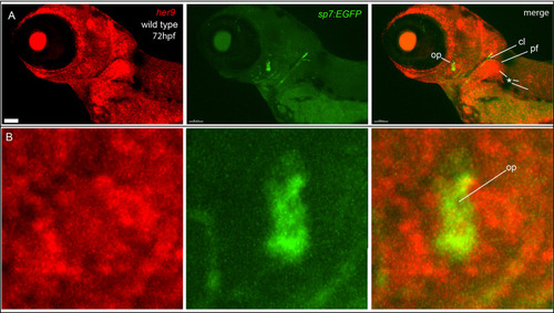
her9 is broadly expressed, but not enriched in osteoblasts. (A) In situ hybridization detects broad her9 expression at 72 hpf. Asterisks indicate areas of strong her9 expression. Opercle osteoblasts do not strongly express her9. (B) Enlargements of images in (A). Images in (B) are enlargements of (A). Scale bar is 80 μm. EXPRESSION / LABELING:
|
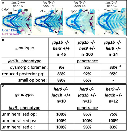
Mutations in her9 enhance jag1b mutant phenotypes. (A) jag1b;her9 double heterozygotes were intercrossed and six days post fertilization (dpf) larvae were stained with Alcian Blue and Alizarin Red to label cartilage and bone. The individuals were then genotyped, the viscerocrania were dissected and flat mounted then imaged. The following craniofacial skeletal elements are indicated in the wild-type individual: opercle bone (op), branchiostegal ray (br), cleithrum (cl), foramen (f) palatoquadrate (pq). The dysmorphic foramen (df) and reduced posterior pq (rppq) phenotypes are indicated, and the small op bone phenotype is indicated by an arrowhead. Arrows mark the opercle mineralization defect associated with her9 homozygous mutants. Poorly stained posterior arch derived cartilages and one missing entopterygoid bone in jag1b-/-;her9+/- are staining and mounting artifacts in this individual and not representative phenotypes associated with this genotype. Scale bar is 50 μm (B) Genotyped preps from A were scored for penetrance of jag1b mutant-associated phenotypes. Asterisk in dysmorphic foramen row indicates that jag1b-/-;her9-/- is significantly different from both jag1b-/-;her9+/+ and jag1b-/-;her9+/- using a Fisher’s exact test with p<0.05. (C) Genotyped preps from A were scored for penetrance of her9 mutant-associated phenotypes. PHENOTYPE:
|

Mutations in her6 enhance the jag1b mutant phenotype, her6 and her9 are redundantly required for eye development. (A) Exon diagram for her6 indicating the untranslated regions (UTR) and the regions predicted to encode the basic helix-loop-helix (bHLH) and Orange domains. The location of the deletion encoded by the her6co3005 allele is indicated. (B) jag1b;her6 double heterozygotes were intercrossed and six days post fertilization (dpf) larvae were stained with Alcian Blue and Alizarin Red to label cartilage and bone. The individuals were then genotyped, the viscerocrania were dissected and flat mounted then imaged. The following craniofacial skeletal elements are indicated in the wild-type individual: opercle bone (op), branchiostegal ray (br), cleithrum (cl), foramen (f). The dysmorphic foramen phenotype (df) is indicated, and the small op bone phenotype is indicated by an arrowhead. Scale bar is 50 μm (C) Genotyped preps from B were scored for penetrance of jag1b mutant-associated phenotypes. Asterisk in small op bone row indicates that penetrance in jag1b-/-;her6-/- is significantly different from jag1b-/-;her6+/+ (p<0.05). (D) her6;her9 double heterozygotes were intercrossed and six days post fertilization (dpf) larvae were stained with Alcian Blue and Alizarin Red to label cartilage and bone. The individuals were then genotyped and whole-mount imaged. An arrow indicates unmineralized opercle. The lens (l) is indicated in the wild type and a double lens phenotype is indicated in double homozygous mutants (dl). PHENOTYPE:
|
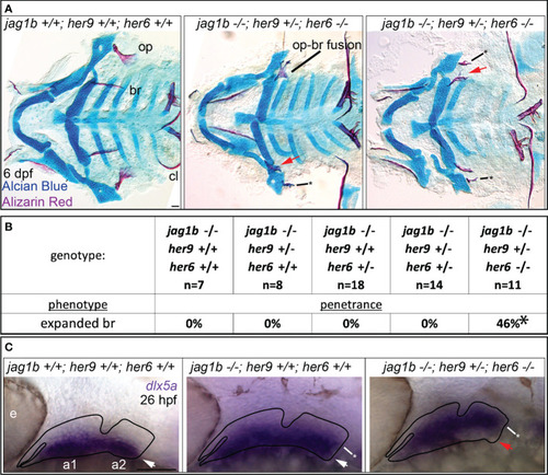
jag1b, her9, and her6 redundantly regulate dorsal versus ventral identity. (A) jag1b;her6;her9 triple heterozygotes were intercrossed and six days post fertilization (dpf) larvae were stained with Alcian Blue and Alizarin Red to label cartilage and bone. The individuals were then genotyped, the viscerocrania were dissected and flat mounted then imaged. The opercle (op), branchiostegal ray (br), and cleithrum (cl) are indicated in the wild type control. The bony fusion between the opercle and the branchiostegal ray is indicated. Red arrows indicate expanded, fan-shaped branchiostegal ray. Asterisk marks stick-shaped op phenotype (B) Genotyped preps from A were scored for penetrance of expanded branchiostegal ray. Asterisk indicates that penetrance of this phenotype in jag1b-/-;her9+/-;her6-/- is significantly different from all other genotypes, where it is never observed. (C) jag1b;her6;her9 triple heterozygotes were intercrossed and 26 hours post fertilization (hpf) larvae were fixed for in situ hybridization experiments to label dlx5a expression. White arrows indicate ventral dlx5a expression, asterisks mark dorsally expanded dlx5a expression. Red arrow indicates reduced ventral dlx5a expression in triple mutants suggesting ventral-versus-dorsal identity reversal. This phenotype was observed on both sides of in 3/3 embryos examined with this genotype. Scale bars are 50 μm. |

ZFIN is incorporating published figure images and captions as part of an ongoing project. Figures from some publications have not yet been curated, or are not available for display because of copyright restrictions. |
|
ZFIN is incorporating published figure images and captions as part of an ongoing project. Figures from some publications have not yet been curated, or are not available for display because of copyright restrictions. PHENOTYPE:
|