
FIGURE
Figure 5
- ID
- ZDB-FIG-221230-14
- Publication
- Stenzel et al., 2022 - Distinct and redundant roles for zebrafish her genes during mineralization and craniofacial patterning
- Other Figures
- All Figure Page
- Back to All Figure Page
Figure 5
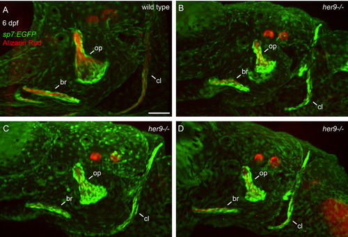
Opercle shapes are variable in her9 mutants. sp7:EGFP;her9 heterozygotes were crossed to her9 heterozygotes and offspring were sorted for transgene expression. 24 live transgenic animals were labeled with Alizarin Red and imaged at 6 dpf. Imaged animals were genotyped. (A) Representative opercle region from a wild-type larva. (B–D) Three different her9 mutant larvae are shown. The following structures are indicated: opercle bone (op), branchiostegal ray (br), cleithrum (cl). Scale bar is 50 μm. |
Expression Data
Expression Detail
Antibody Labeling
Phenotype Data
| Fish: | |
|---|---|
| Observed In: | |
| Stage: | Day 6 |
Phenotype Detail
Acknowledgments
This image is the copyrighted work of the attributed author or publisher, and
ZFIN has permission only to display this image to its users.
Additional permissions should be obtained from the applicable author or publisher of the image.
Full text @ Front Endocrinol (Lausanne)