
- Title
-
Genetic duplication of tissue factor reveals subfunctionalization in venous and arterial hemostasis
- Authors
- Grzegorski, S.J., Zhao, Y., Richter, C.E., Ku, C.J., Lavik, K.I., Paul, D., Morrissey, J.H., Shavit, J.A.
- Source
- Full text @ PLoS Genet.
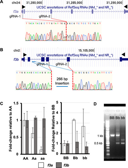
Genome engineering creates large loss-of-function deletions in both copies of zebrafish f3.
Two sgRNAs each for f3a and f3b were complexed with Cas9 and injected into single cell embryos. (A) A large deletion was identified between exon 1 and exon 3 in f3a resulting in a nonsense mutation. (B) A deletion was introduced between exon 4 and exon 5 of f3b. During endogenous repair, a section of intronic DNA (blue bar) was inverted and inserted at the site of the deletion introducing a premature stop codon. Black arrowheads indicate the locations of primers used for full length cDNA amplification. (C) RT-qPCR at 4 dpf demonstrated that the mutant f3a allele resulted in lower mRNA levels (consistent with nonsense mediated decay of the residual transcript) without affecting f3b levels. Conversely, loss of f3b induces upregulation of f3a, but the levels of f3b transcript were not diminished. (D) f3b cDNA was isolated from pools of 4 dpf larvae from heterozygous mutant incrosses. The expected wild-type band was visible in the BB and Bb mutants. Two smaller bands were evident in the Bb and bb pools, consistent with alternative splicing of the mutant transcript. |
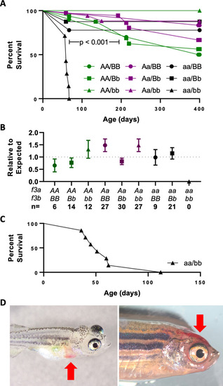
Complete loss of TF activity results in early lethality due to hemorrhage.
(A) Fish from a double heterozygous (Aa/Bb) f3 incross were genotyped at 8 weeks of age and found to have normal Mendelian ratios (n = 135, AA/BB = 10, AA/Bb = 17, AA/bb = 7, Aa/BB = 15, Aa/Bb = 36, Aa/bb = 20, aa/BB = 10, aa/Bb = 13, aa/bb = 9). Survival curves show that by 9 weeks, there was statistically significant loss of aa/bb offspring. By 400 days, there was a relatively even reduction of the remaining genotypes which is common in wild-type fish, and confirms that a single copy of f3a or f3b is sufficient for survival. (B) A repeat cohort was left undisturbed until genotyping at 6 months of age. The statistically significant loss of aa/bb confirms the early lethality of TF loss independent of genotyping stress. (C) A group of 7 double homozygous mutants were identified at 1 month of age and observed over the following 3 months. 6/7 were lost by 2 months with the single survivor passing at 112 days. (D) Early lethality was due to gross hemorrhage. Examples include the pericardial space (left) and head (right) (red arrows). |
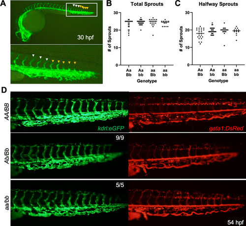
Vascular development is unaffected in the absence of TF.
(A) Example of a 30 hpf larva carrying the kdrl:eGFP transgene marking vascular endothelial cells (top) with a close-up (below). White arrowheads mark examples of vessel sprouts that have made it at least halfway through the expected migration. Orange arrowheads represent sprouts that have not yet passed the halfway point. No statistically significant changes were observed between groups in total vessel sprouts (B) or sprouts that have made it past the halfway point (C) by Mann-Whitney U testing. (D) At 54 hpf, no observable changes are seen in endothelial cell development (left, green) or in vascular integrity (right, red). The latter is evidenced by long exposure in the gata1:DsRed transgenic background in which erythrocytes are marked by red fluorescence. Representative images for 14 experimental mutant larvae that were scored by a blinded observer are shown along with a wild type control. |
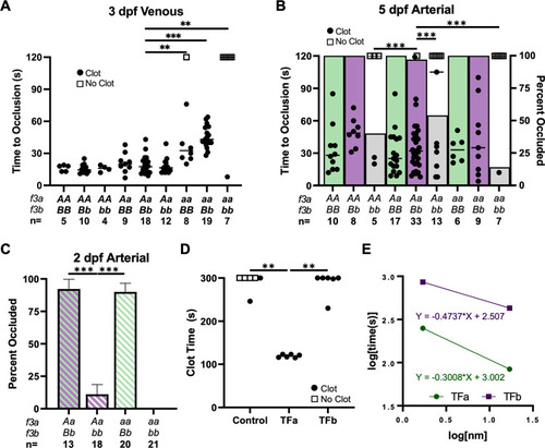
TFa and TFb have evolved partial subfunctionalization according to in vivo vascular endothelial laser injury model and in vitro recombinant protein clotting assays.
(A) Laser-mediated venous endothelial injury at 3 dpf shows that the time to occlusion (TTO) was not altered by loss of TFb (AA/bb, Aa/bb) but was delayed in the complete absence of TFa (aa/BB, aa/Bb). Thrombus formation was absent after complete TF loss (aa/bb). (B) Laser-mediated arterial endothelial injury at 5 dpf shows that the ability to form occlusive thrombi was significantly reduced in fish lacking TFb (AA/bb, Aa/bb, aa/bb, indicated by grey bars) compared to BB and Bb groups (green and purple bars; respectively). (C) Endothelial injury at 2 dpf confirms the dependence on TFb in the arterial system prior to the presence of thrombocytes. Occlusive thrombi were nearly absent without TFb (Aa/bb, aa/bb). (D) Relipidated recombinant TFa or TFb proteins (173 nM) were mixed with citrated trout plasma, and recalcified. TFa-liposomes induced clot formation during a manual tilt test significantly faster than TFb-liposomes or empty liposome controls. The reaction was observed until 300 seconds, and then again at 15 minutes to assess for delayed clot formation (the latter observation is indicated by an open or closed circle at 300 seconds). (E) The assay was repeated with relipidated TF diluted in 0.5x HBS (20 mM HEPES pH 7.5, 50 mM NaCl) and plotted on log-log axes demonstrating 10 to 100-fold higher procoagulant activity of TFa over TFb. Statistical significance by Mann-Whitney U testing: ** < 0.01, *** < 0.001. All phenotypic data were collected by an observer blinded to genotype. Open squares in A and B indicate individual larvae that did not occlude within 120 seconds. |
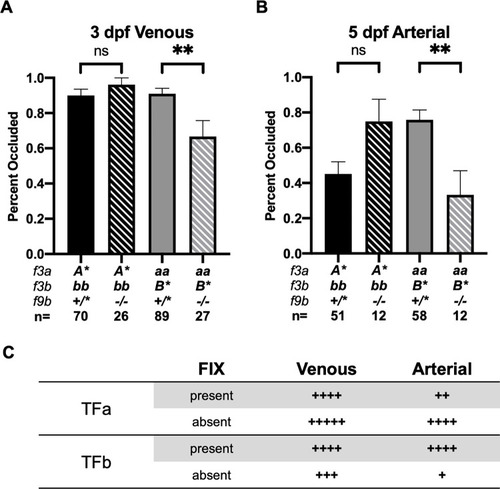
Loss of FIXb impairs clot formation in f3a-null offspring.
(A) Loss of f9b does not influence induced venous occlusion in A*/bb but does decrease total frequency of occlusion in aa/B* larvae at 3 dpf. (B) Loss of f9b does not significantly affect clot formation in A*/bb offspring. In aa/B* larvae, loss of f9b leads to a significant impairment of occlusive clot formation. Statistical significance by Mann-Whitney U testing: ** < 0.01. A* represents AA or Aa, B* represents BB or Bb. (C) Summary of data in (A) and (B) with proposed relative ohnolog occlusive activity in the presence and absence of FIX, in arterial versus venous systems. |
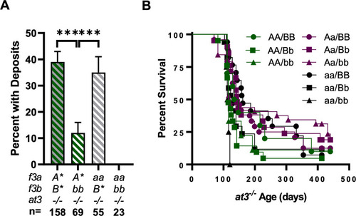
Loss of TFb protects against spontaneous thrombosis in at3-/- larvae but does not improve long term survival.
(A) Fibrin deposits were assessed at 5 dpf. Deficiency of TFa did not alter thrombosis scores while loss of TFb led to less deposition (B) When followed for >1 year, f3 genotype did not influence at3-/- survival; however, all aa/bb offspring were lost by 120 days, consistent with TF dependent lethality. Statistical significance by binomial proportion test: *** < 0.001. A* represents AA or Aa, B* represents BB or Bb. |
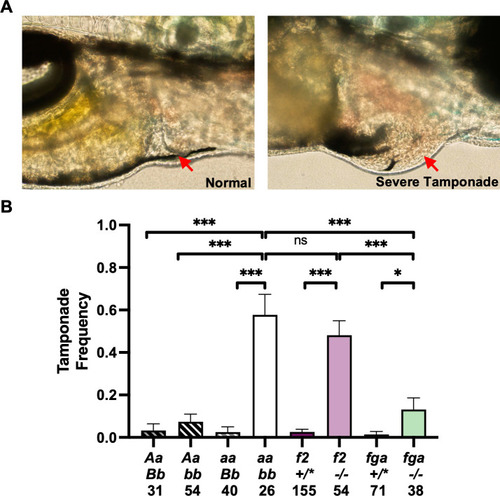
Loss of TF contributes to risk of cardiac tamponade.
(A) Stress was chemically induced in 3 dpf larvae by exposure for 16 hours using 25 μM epinephrine and 0.01% hydrocortisone. Larvae were scored for the presence of erythrocytes in the pericardial space (noted by red arrows) by an observer blinded to genotype. Examples of control (left) and tamponade-positive fish (right) are shown. (B) TF-, prothrombin (f2)- and fibrinogen (fga)-deficient fish all showed significantly elevated rates of tamponade relative to controls. Furthermore, TF- and prothrombin-deficient larvae had comparable rates of tamponade and are both significantly increased relative to fga mutants. Statistical significance by binomial proportion test: * < 0.5, ** < 0.01, *** < 0.001. +/* represents +/+ or +/-. |