FIGURE SUMMARY
Title

Biliverdin regulates NR2E3 and zebrafish retinal photoreceptor development

Authors
Connor, B., Titialii-Torres, K., Rockenhaus, A.E., Passamonte, S., Morris, A.C., Lee, Y.S.
Source
Full text @ Sci. Rep.

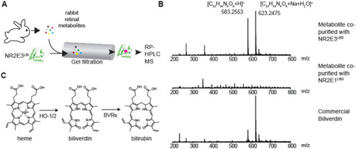
Identification of biliverdin as a potential NR2E3LBD ligand. (A) A schematic representation of the method. Small molecules extracted from rabbit retina were incubated with recombinant MBP-NR2E3LBD expressed and purified from E. coli. Protein-bound small molecules were isolated by a gel filtration chromatography and identified by mass spectrometry. (B) Mass spectra (ESI, positive ions) of metabolites co-purified with MBP-NR2E3LBD, ones eluted in the gel filtration fraction without any protein, and commercial biliverdin. (C) A schematic representation of biliverdin and its metabolism (HO-1/2: heme oxygenases 1 and 2, BVRs: biliverdin reductases).

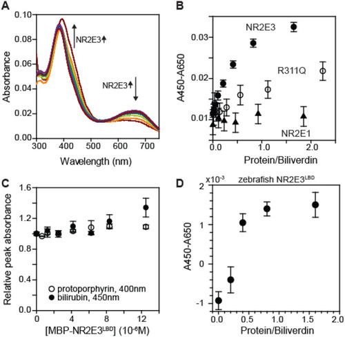
Biliverdin specifically binds to MBP-NR2E3LBD in vitro. (A) Visible spectra of biliverdin (5 μM) in the absence and in the presence of varying amounts of MBP-NR2E3LBD. The blue peak shifts from 390 to 450 nm, while the red peak intensity diminishes upon binding to MBP-NR2E3LBD. (B) Biliverdin stoichiometrically bound to MBP-NR2E3LBD (filled circle). MBP-NR2E3LBD R311Q variant (open circle) showed a weaker binding to biliverdin (5 μM). MBP-NR2E1LBD (triangle) did not show any evidence of binding to biliverdin. Averages and standard deviations (n = 3) are presented. (C) Effect of MBP-NR2E3LBD on visible spectra of (filled circle) bilirubin and (open circle) protoporphyrin IX (average and standard deviations, n = 3). (D) Effect of MBP-zebrafish NR2E3LBD to the visible spectrum of biliverdin (average and standard deviations, n = 3). The concentration of biliverdin was identical to the one used in Fig. 2A, B.

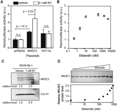
Biliverdin regulates NR2E3 in cells. (A, B) Reporter gene expression analysis in 293F cells. Cells were transfected with a reporter plasmid encoding nanoluciferase under the control of 9 × GALUAS and plasmids (pFN26A derivatives) encoding GALDBD-NR2E3 fusion protein. A day after transfection, cells were treated with vehicle (0.1% DMSO) or biliverdin for 3 h. Nanoluciferase activity was measured using Vivazine substrate (average and standard deviations, n = 3). (C, D) Biliverdin increased NR2E3 protein level in WERI-Rb-1 cells. WERI-Rb-1 human retinoblastoma cells were incubated with vehicle (0.1% DMSO) or biliverdin (1 μM for C and varying concentrations for D) for 24 h. Western blot results (1:1000-fold diluted antibody) are shown. COX IV was used as a loading control. In Fig. 3C, different parts of the same gel are shown. Full-sized Western blot images are available in the Supplementary Information.

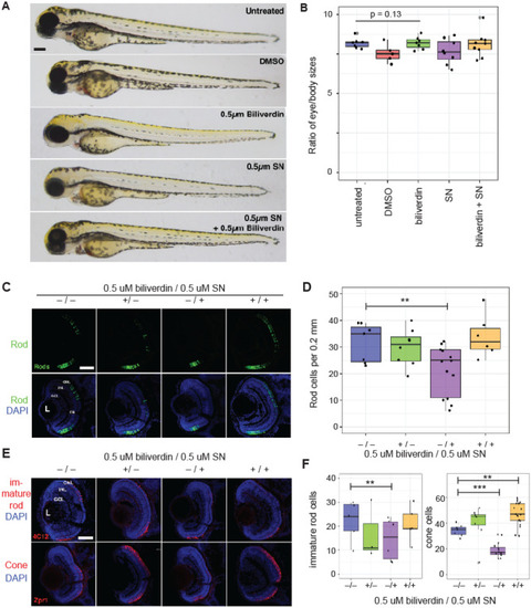
Biliverdin contributes to retinal development in zebrafish larvae. (A, B) Biliverdin and sulconazole at the tested concentrations (0.5 μM each) did not affect the overall development of zebrafish larvae. (A) Whole mount images of 3 dpf zebrafish larvae. (B) Quantification of the eye and body size ratios. (C) Fluorescence microscopic images of 3 dpf zebrafish larvae (from left to right) without any treatment, with 0.5 μM biliverdin, with 0.5 μM sulconazole nitrate, and with both biliverdin and sulconazole nitrate (top: rod photoreceptor cells, bottom: overlay of rod photoreceptor cells and DAPI-stained cells). (D) Quantification of rod photoreceptor cells in zebrafish larvae (3 dpf) grown in different conditions (**p = 0.00369). (E) Fluorescence microscopic images of zebrafish larvae (top) immunolabeled with the 4C12 antibody (which detect immature rod photoreceptors) and (bottom) red-green cones immunolabeled with the Zpr-1 antibody. Nuclei were counterstained with DAPI. (F) Quantification of immature rods and red-green cones (***p < 10−5). ONL, outer nuclear layer; INL, inner nuclear layer; GCL, ganglion cell layer. Scale bars, 0.1 mm in A; 0.05 mm in C, E.

Acknowledgments
This image is the copyrighted work of the attributed author or publisher, and ZFIN has permission only to display this image to its users. Additional permissions should be obtained from the applicable author or publisher of the image. Full text @ Sci. Rep.