IMAGE

Figure 4

ID
ZDB-IMAGE-220509-25
Antibodies
Source
Figures for Connor et al., 2022
Image
Figure Caption

Figure 4

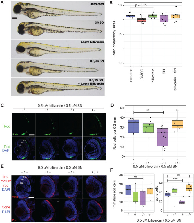
Biliverdin contributes to retinal development in zebrafish larvae. (A, B) Biliverdin and sulconazole at the tested concentrations (0.5 μM each) did not affect the overall development of zebrafish larvae. (A) Whole mount images of 3 dpf zebrafish larvae. (B) Quantification of the eye and body size ratios. (C) Fluorescence microscopic images of 3 dpf zebrafish larvae (from left to right) without any treatment, with 0.5 μM biliverdin, with 0.5 μM sulconazole nitrate, and with both biliverdin and sulconazole nitrate (top: rod photoreceptor cells, bottom: overlay of rod photoreceptor cells and DAPI-stained cells). (D) Quantification of rod photoreceptor cells in zebrafish larvae (3 dpf) grown in different conditions (**p = 0.00369). (E) Fluorescence microscopic images of zebrafish larvae (top) immunolabeled with the 4C12 antibody (which detect immature rod photoreceptors) and (bottom) red-green cones immunolabeled with the Zpr-1 antibody. Nuclei were counterstained with DAPI. (F) Quantification of immature rods and red-green cones (***p < 10−5). ONL, outer nuclear layer; INL, inner nuclear layer; GCL, ganglion cell layer. Scale bars, 0.1 mm in A; 0.05 mm in C, E.

Figure Data
Acknowledgments
This image is the copyrighted work of the attributed author or publisher, and ZFIN has permission only to display this image to its users. Additional permissions should be obtained from the applicable author or publisher of the image. Full text @ Sci. Rep.