
- Title
-
A Novel Missense Variant in Actin Binding Domain of MYH7 Is Associated With Left Ventricular Noncompaction
- Authors
- Hesaraki, M., Bora, U., Pahlavan, S., Salehi, N., Mousavi, S.A., Barekat, M., Rasouli, S.J., Baharvand, H., Ozhan, G., Totonchi, M.
- Source
- Full text @ Front Cardiovasc Med

ZFIN is incorporating published figure images and captions as part of an ongoing project. Figures from some publications have not yet been curated, or are not available for display because of copyright restrictions. PHENOTYPE:
|
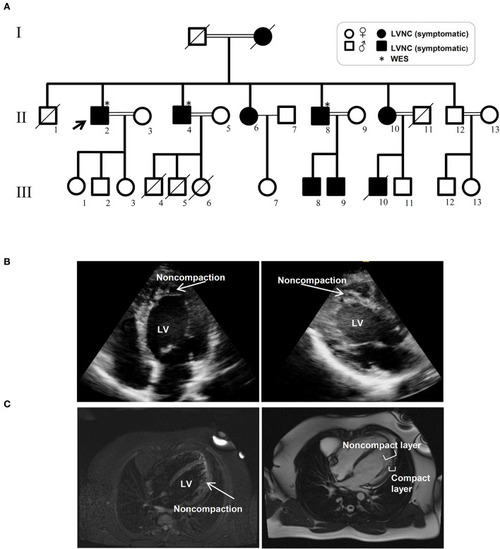
Pedigree and clinical manifestation of the left ventricular non-compaction (LVNC) in the affected members included in this study. |
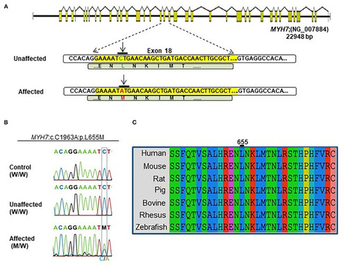
Identification of the |
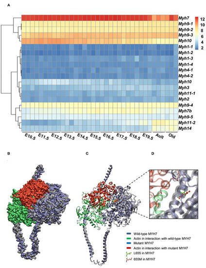
Expression patterns of all |
The EXPRESSION / LABELING:
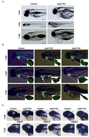
PHENOTYPE:
|