
- Title
-
The Abcc6a Knockout Zebrafish Model as a Novel Tool for Drug Screening for Pseudoxanthoma Elasticum
- Authors
- Van Gils, M., Willaert, A., Coucke, P.J., Vanakker, O.M.
- Source
- Full text @ Front Pharmacol
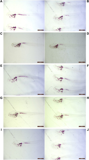
Light microscopy examples of ARS-stained abcc6acmg52/cmg52 specimens. Examples of 10 dpf untreated PHENOTYPE:
|
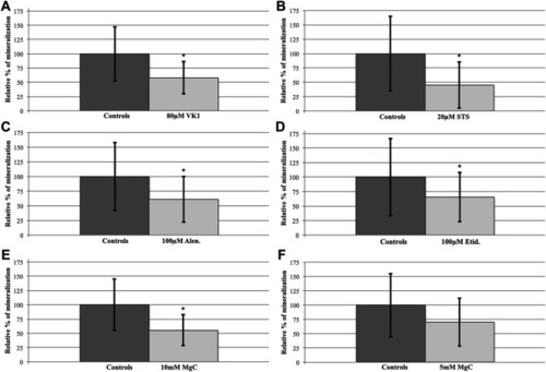
Compound treatment effects on the abcc6acmg52/cmg52 spinal hypermineralization phenotype. After semiquantification and statistical analysis, mean ± SD percentile values of the spinal mineralization were normalized per compound to the respective control values of the untreated abcc6acmg52/cmg52 fish. |
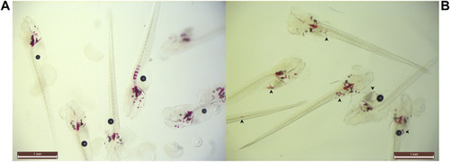
Example of ectopic speckling following 35 µM STS-treatment. Snapshot group images of PHENOTYPE:
|
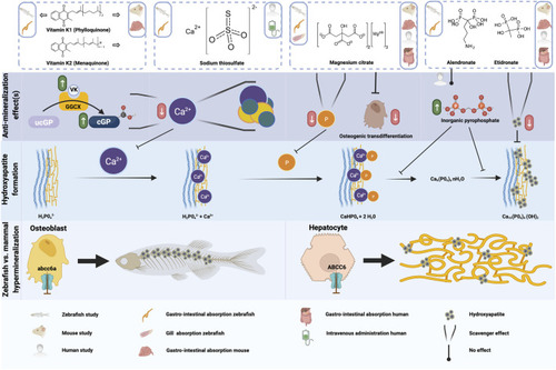
Schematic representation of the anti-mineralization effects of the tested compounds. For the compounds evaluated in the present study, the type of administration or absorption is mentioned in the top row, as well as the type of administration used in any mouse or human studies which were done with this compound. The anti mineralizing effects of the different compounds are shown, as well as where these effect take place in the cascade to form hydroxyapatite crystals. Finally, the differences between the hypermineralization in zebrafish and mammals with respect to abcc6a and ABCC6 deficiency are shown. Figure created with |