
- Title
-
brca2-mutant zebrafish exhibit context- and tissue-dependent alterations in cell phenotypes and response to injury
- Authors
- Kouprianov, V.A., Selmek, A.A., Ferguson, J.L., Mo, X., Shive, H.R.
- Source
- Full text @ Sci. Rep.
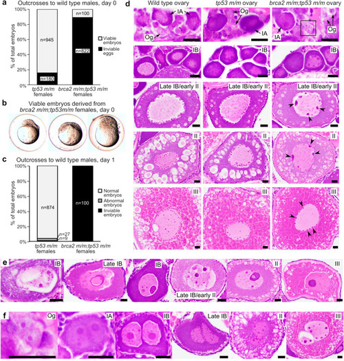
Embryos from brca2 m/m;tp53 m/m female zebrafish undergo proliferation arrest and death and oocytes exhibit abnormal nuclear morphology. (a) Most eggs derived from brca2 m/m;tp53 m/m females are inviable and only a small number of viable embryos are generated, in contrast to tp53 m/m females. Females were outcrossed to fertile wild type males. (b) On day 0, viable embryos derived from brca2 m/m;tp53 m/m embryos are often morphologically abnormal and all undergo developmental arrest at or before sphere stage (approximately four hours post-fertilization). (c) At one day post-fertilization (day 1), greater than 90% of embryos derived from tp53 m/m females are alive and morphologically normal, while all embryos derived from brca2 m/m;tp53 m/m females are dead. (d) Oocytes from brca2 m/m;tp53 m/m females exhibit nuclear abnormalities predominated by aggregated nucleolar material around nuclear margins (black arrowheads). Black box indicates oogonium shown at higher magnification in panel f. (e) Oocytes with massive nucleolar condensation are often degenerate. (f) Infrequent binucleation occurred in oogonia and oocytes from brca2 m/m;tp53 m/m females. Og, oogonia; IA, stage IA oocyte; IB, stage IB oocyte; II, stage II oocyte; III, stage III oocyte. Scale bar = 20 µm. |
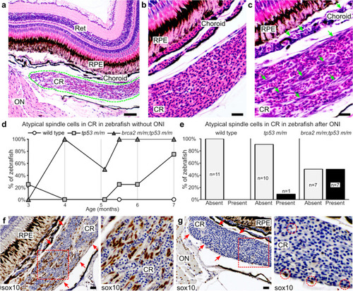
Atypical spindle cells accumulate in the choroid rete of brca2 m/m;tp53 m/m zebrafish. (a) The choroid rete (outlined in green) is a vascular plexus located subjacent to the choroid. (b) Normal choroid rete. (c) Choriod rete containing numerous atypical spindle cells (green arrows). (d) Numbers of zebrafish that developed atypical spindle cells in the choroid rete over time (n = 4 per genotype at each time point analyzed). (e) Numbers of zebrafish that received optic nerve injury and developed atypical spindle cells in the choroid rete. (f) Atypical spindle cells in the choroid rete are sox10-positive (brown chromogen). Red arrows delineate margins of choroid rete. Area boxed in red is shown at higher magnification to the right. (g) Small numbers of sox10-positive cells (brown chromogen) are present in the normal choroid rete. Red arrows delineate margins of choroid rete. Area boxed in red is shown at higher magnification to the right. Red circles identify sox10-positive cells. Ret, retina; RPE, retinal pigmented epithelium; CR, choroid rete; ON, optic nerve. Scale bar = 50 µm (panel a); 20 µm (panels b, c, f, h). |
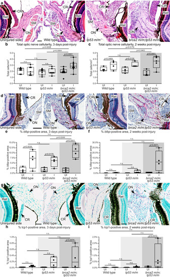
brca2 m/m;tp53 m/m zebrafish exhibit increased proliferative responses in the injured optic nerve. (a) Representative histologic section of the optic nerve pathway after unilateral optic nerve injury. (b,c) Quantitative analysis of the total cellularity in the uninjured versus injured optic nerve three days (b) and two weeks (c) post-injury. (d) Representative examples of blbp expression (red chromogen), a marker for radial glial cells, in the uninjured and injured optic nerves. (e,f) Quantitative analysis of blbp-positive area in the uninjured versus injured optic nerve three days (e) and two weeks (f) post-injury. (g) Representative examples of lcp1 expression (purple chromogen), a marker for monocytes/macrophages, in the uninjured and injured optic nerves. (h,i) Quantitative analysis of lcp1-positive area in the uninjured versus injured optic nerve three days (h) and two weeks (i) post-injury. Ret, retina; SM, skeletal muscle; CR, choroid rete; ON, optic nerve; UI, uninjured; I, injured. Scale bar = 50 µm (panels a, g); 100 µm (panel d). All images depict specimens collected at three days post-injury except panel g, in which the brca2 m/m;tp53 m/m specimen shown was collected at two weeks post-injury. |
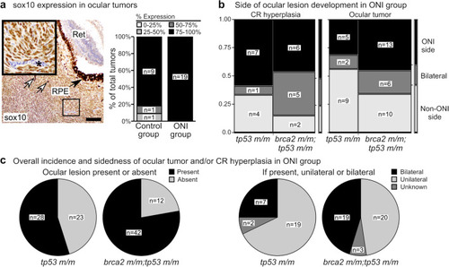
Optic nerve injury (ONI) and brca2 genotype exert variable effects on the development of proliferative and neoplastic lesions in the optic nerve pathway. (a) The majority of zebrafish ocular tumors highly express sox10 regardless of ONI status. White arrows indicate dispersed melanin pigment within tumors. Inset shows tumor cells with positive nuclear sox10 expression (brown chromogen). Asterisk indicates a blood vessel containing nucleated erythrocytes that do not express sox10. (b) In zebrafish that received ONI, most brca2 m/m;tp53 m/m zebrafish developed atypical spindle cells in the choroid rete (CR) on the injured side (ONI side) or bilaterally, in contrast to ocular tumor development. (c) In zebrafish that received ONI, brca2 m/m;tp53m/m zebrafish were more likely to develop ocular lesions (ocular tumor and/or hyperplastic spindle cells in the choroid rete) and these lesions were more likely to be bilateral. HE, hematoxylin and eosin; Ret, retina; RPE, retinal pigmented epithelium; ONI, optic nerve injury; CR, choroid rete. Scale bar = 50 µm. PHENOTYPE:
|