
- Title
-
Pathogenic effect of TP73 Gene Variants in People With Amyotrophic Lateral Sclerosis
- Authors
- Russell, K.L., Downie, J.M., Gibson, S.B., Tsetsou, S., Keefe, M.D., Duran, J.A., Figueroa, K.P., Bromberg, M.B., Murtaugh, L.C., Bonkowsky, J.L., Pulst, S.M., Jorde, L.B.
- Source
- Full text @ Neurology
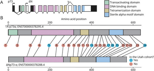
Twenty-Four Rare (ExAC NFE MAF <0.001) TP73 Nonsynonymous Variants Were Found Across Multiple Cohorts (A) TA-p73 and ΔN-p73, which have opposing tumor suppressive/oncogenic functions, are expressed from 2 separate promoters. Alternative splicing events also give rise to a multitude of different isoforms (such as ΔN-p73α and ΔN-p73γ). (B) Primary structure of TA-p73α and ΔN-p73α. Number within the circles refers to the variant number in table 1. ExAC = Exome Aggregation Consortium; MAF = minor allele frequency; NFE = non-Finnish European. |
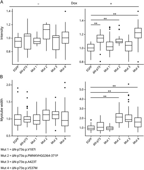
ALS Variants in TP73 Render p73 Dysfunctional in C2C12 Differentiation Demonstrated by Quantification of MHC Staining Intensity and Myotube Width Compared to EGFP (A) Quantification of normalized myosin heavy chain (MHC) staining in differentiated C2C12s, with or without exposure to doxycycline, expressing enhanced green fluorescent protein (EGFP), wild-type (WT) ΔN-p73α, or mutant ΔN-p73. WT ΔN-p73α expression results in inhibited C2C12 differentiation shown by reduced MHC immunohistochemistry staining compared to EGFP. ΔN-p73αMut1+3 expression fails to inhibit C1C12 differentiation shown by no difference in MHC staining compared to EGFP (**p < 0.001, Wilcoxon rank-sum test with Bonferroni correction). (B) Normalized myotube width quantification. ΔN-p73αMut2-4 expression results in myotubes with significantly larger width compared to EGFP. ALS = amyotrophic lateral sclerosis. |
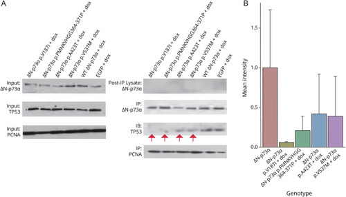
ΔN-p73α Containing ALS Patient Mutations Expressed in Neuro-2A Cells Displayed Impeded Binding to Proapoptotic p53 (A) Western blots show equal input level of protein (proliferating cell nuclear antigen [PCNA], ΔN-p73α, and p53) before immunoprecipitation. Protein was immunoprecipitated with ΔN-p73α or PCNA antibody. Postimmunoprecipitation lysate demonstrates that all ΔN-p73α protein was immunoprecipitated with ΔN-p73α antibody. Blots were probed for ΔN-p73α, p53, and PCNA. PCNA was used as a loading control and was immunoprecipitated in tandem to ΔN-p73α. Red arrows denote less intense p53 protein bands immunoprecipitated by mutant ΔN-p73α compared to wild-type (WT) ΔN-p73α and enhanced green fluorescent protein (EGFP) (in EGFP lane, ΔN-p73α band is endogenous protein). (B) Quantification of p53 band intensity of Western blots performed after ΔN-p73α immunoprecipitation. Shown are mean intensity and SD for p53 bands from coimmunoprecipitations performed in biological duplicate for Neuro-2A cells expressing ST ΔN-p73α or mutant ΔN-p73α on doxycycline (dox) exposure. ALS = amyotrophic lateral sclerosis. |
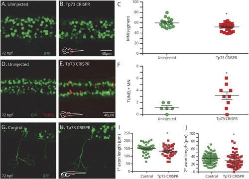
tp73 Mutation Decreases Motor Neuron Count, Increases Apoptosis, and Impairs Motor Neuron Axon Outgrowth (A and B) Confocal images of spinal motor neurons (MNs; green) in uninjected and tp73CRISPR injected embryos (confocal imaging of dorsal view of spinal cord). (C) tp73CRISPR-injected embryos have a significantly reduced number of MNs compared to both uninjected and tyrCRISPR-injected sibling controls (supplement figure 4, C and D, doi:10.5061/dryad.4qrfj6q94). (D and E) Confocal images of increased MN (green) apoptosis (red terminal deoxynucleotidyl transferase dUTP nick-end labeling [TUNEL]) in tp73CRISPR embryos compared to uninjected. (F) Increased MN apoptosis in tp73CRISPR embryos compared to uninjected, performed in mnx:GFP transgenic line. (G and H) Confocal images of MN primary and secondary axons (green) in uninjected and tp73CRISPR-injected embryos. (I) MN primary axon length in tp73CRISPR embryos is significantly shorter than that of uninjected sibling controls. (J) MN secondary axon length in tp73CRISPR-injected embryos is significantly shorter compared to that of uninjected sibling controls. CRISPER = clustered regularly interspaced short palindromic repeats; hpf = high-power field. |