FIGURE SUMMARY
Title

Endogenous zebrafish proneural Cre drivers generated by CRISPR/Cas9 short homology directed targeted integration

Authors
Almeida, M.P., Welker, J.M., Siddiqui, S., Luiken, J., Ekker, S.C., Clark, K.J., Essner, J.J., McGrail, M.
Source
Full text @ Sci. Rep.

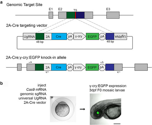
CRISPR/Cas9 short homology directed targeted integration strategy for efficient recovery of Cre knock-in alleles. (a) Schematic of the genomic target site, Cre donor vector, and final knock in allele. A CRISPR sgRNA (red) was chosen in the coding sequence. 48 bp upstream (dark green) and 48 bp downstream (dark blue) of the cut site are included in the donor vector as homology arms. The donor vector has UgRNA sequences flanking the homology arms for in vivo liberation of the knock in cassette. The knock-in cassette contains an in frame 2A-Cre for expression of Cre recombinase, and a secondary marker driving γ-cry:EGFP expression in the lens. Arrows indicate PCR primers for 5′ and 3′ genome/vector junction analysis. (b) 1 cell stage zebrafish embryos are injected with Cas9 mRNA (300 pg), genomic sgRNA (25 pg), UgRNA (25 pg), and 2A-Cre targeting vector (10 pg). At 3 days post fertilization, embryos expressing EGFP in the lens are selected and raised to adulthood. TS genomic sgRNA target site, UgRNA universal short guide RNA target site, γ-cry gamma crystallin promoter, EGFP green fluorescent protein, pA transcription termination and polyadenylation sequence.

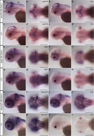
Expression of 2A-Cre integration alleles recapitulates
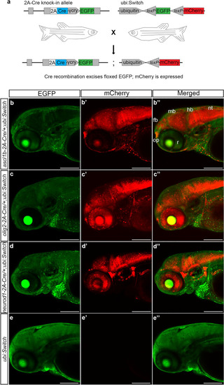
 ascl1b, olig2, and neurod1 expression pattern. Whole mount in situ hybridization for endogenous genes and Cre was performed in 3 dpf larvae obtained from outcrossing ascl1b-2A-Cre/+, olig2-2A-Cre/+, and neurod1-2A-Cre/+ lines to wild type WIK. Sibling larvae from the same clutch were sorted into lens EGFP-positive and -negative groups before fixation. For each genotype and probe 3 individual larvae were photographed. (ad) ascl1b and Cre expression in ascl1b-2A-Cre/+ larvae show similar patterns in the forebrain, and along the midbrain ventricle and midbrain-hindbrain border. (eg) olig2 and Cre expression in olig2-2A-Cre/+ larvae were restricted to the forebrain, the posterior half of the cerebellum, and a subset of cells in the retina. (ik) neurod1 and Cre expression in neurod1-2A-Cre/+ larvae were detected in the forebrain, adjacent to the midbrain and hindbrain ventricles and enriched in the cerebellum ((i′), C black arrow), and in the retina inner (large bracket) and outer (small bracket) nuclear layers (j). neurod1 and Cre expression were not detected in the posterior peripheral midbrain layer ((i′), PML white arrow). Cre expression was not detected in wild type +/+ sibling larvae (d,h,l). c cerebellum, fb forebrain, hb hindbrain, mb midbrain, mhb midbrain-hindbrain border, nt neural tube, ot optic tectum, pml peripheral midbrain layer, r retina. Scale bar 250 μm.

Transgenic ascl1b-2A-Cre, olig2-2A-Cre and neurod1-2A-Cre lines express functional Cre that promotes recombination at loxP sites in the expected neural progenitor populations. (a) F0 adults harboring precise integration alleles were mated to the recombination reporter line ubi:Switch to generate double transgenic 2A-Cre driver; ubi:Switch embryos. (bd) Confocal imaging of 3 dpf double transgenic ascl1b-2A-Cre; ubi:switch (b), olig2b-2A-Cre; ubi:switch (c), and neurod1-2A-Cre; ubi:switch (d) larvae shows a switch from GFP to mCherry expression in neural cells derived from ascl1b, olig2, and neurod1 progenitors. (e) A switch from GFP to mCherry expression isn’t detected in single transgenic ubi:Switch larvae. fb forebrain, γ-cry gamma crystallin promoter, EGFP green fluorescent protein, hb hindbrain, mb midbrain, nt neural tube, op olfactory placode, r retina. Scale bar 200 μm.

ascl1b-2A-Cre, olig2-2A-Cre and neurod1-2A-Cre Cre recombinase activity in the 3 dpf larval midbrain and hindbrain. F3 or F2 adults were mated to the recombination reporter line ubi:Switch to generate double transgenic embryos. Confocal imaging of ascl1b-2A-Cre; ubi:Switch (aa′′,bb′′), control sibling ubi:Switch (c,d), olig2-2A-Cre; ubi:Switch (ee′′,ff′′) and neurod1-2A-Cre; ubi:Switch (gg′′,hh′′) larval midbrain tectum (t), cerebellum (c) and hindbrain (h). Arrows point to the ventricular zone lining the midbrain and hindbrain ventricles (v). Expression of ascl1b-2A-Cre earlier in brain development leads to nearly all descendant neurons expressing mCherry (a,b). No switching occurs in the absence of Cre (cc′′,dd′′). olig2-2A-Cre leads to mCherry expression in a subset of cells in the cerebellum (arrowheads), and neural progenitors lining the hindbrain ventricular zone and their descendants (e,f). In neurod1-2A-Cre mCherry expression is absent from the ventricular zone (v arrows) but present throughout neurons in the tectum, cerebellum (arrowheads) and hindbrain (g,h). Scale bars 50 μm.

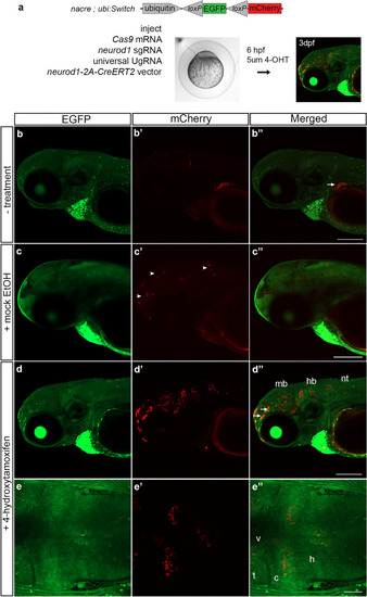
Tamoxifen regulated CreERT2 recombinase activity in F0 somatic targeted embryos. (a) nacre; ubi:Switch embryos injected with Cas9mRNA, sgRNAs and the neurod1-2A-CreERT2 targeting vector. At 6 hpf embryos were mock treated or treated with 5um 4-hydroxytamoxifen and imaged at 3 dpf. (bb′′) Injected embryos without treatment or (cc′′) mock treated show a low level of expression in the brain. (dd′′) 4-hydroxytamoxifen treatment resulted in switching from EGFP to mCherry expression in cells in the midbrain, hindbrain and neural tube. Arrows point to off-target expression. (ee′′) Dorsal imaging of the brain in the larvae shown in dd′′ revealed mCherry expression was detected in cells throughout the midbrain and hindbrain parenchymal tissue. 4-OHT 4-hydroxytamoxifen, c cerebellum, EGFP enhanced green fluorescent protein, hb hindbrain, mb midbrain, nt neural tube, t optic tectum, v ventricle. Scale bars (b′′,c′′,d′′) 200 μm; (e′′) 50 μm.

Acknowledgments
This image is the copyrighted work of the attributed author or publisher, and ZFIN has permission only to display this image to its users. Additional permissions should be obtained from the applicable author or publisher of the image. Full text @ Sci. Rep.