FIGURE SUMMARY
Title

Nrl Is Dispensable for Specification of Rod Photoreceptors in Adult Zebrafish Despite Its Deeply Conserved Requirement Earlier in Ontogeny

Authors
Oel, A.P., Neil, G.J., Dong, E.M., Balay, S.D., Collett, K., Allison, W.T.
Source
Full text @ iScience

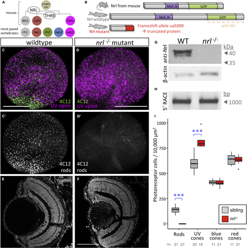
Nrl Is Conserved and Required for Rod Specification in Larval Zebrafish

(A) Nrl is the master regulator of photoreceptor specification in mice, being both necessary and sufficient for rod photoreceptor development from progenitor cells. Elegantly simple models can account for cell fate specification and generating the full complement of photoreceptor types in mice (and all mammals studied) using only two factors, NRL and THRB (thyroid hormone receptor β), to generate rods, red cones, and blue cones expressing RH1, LWS, and SWS opsins, respectively. However, most vertebrates possess additional cone subtypes.

(B) Zebrafish Nrl protein is recognizably similar to mammalian homologs. An nrl−/− null mutation was engineered via CRISPR that is predicted to truncate the protein (see also Figure S1).

(C and D) CRISPR-engineered null Nrl mutants lack rods in larval zebrafish, matching the phenotype of adult NRL−/− knockout mice. Flat-mounted retina from larval zebrafish show that rods are absent in nrl−/− larvae, as detected by rod immunomarker 4C12. The typical broad distribution of rods (B′) is absent (C′) and the decrease in rod cell abundance is consistent, nearly complete and robustly significant (I).

(E and F) Retina of nrl−/− larvae is broadly normal in its lamination.

(G) Nrl protein is not detectable in nrl−/− retina, and antibody validation is supported by a doublet band (presumably reflecting SUMOylation as per mammalian homologs of Nrl) appearing at the predicted size of 44.2 kDa (See Figure S1 for full immunoblots and quantification).

(H) Assessment of nrl transcript by 5′RACE (random amplification of cDNA ends) does not reveal any unexpected transcripts such as those with cryptic exons that could be imagined to make functional protein.

(I) Quantification of photoreceptor types in nrl−/− larvae confirms the consistent absence of rod cells and a concomitant increase in UV cone abundance. Box and whisker plots first through third quartile and distribution of data, respectively, after excluding outliers. ∗∗∗p < 0.001; bar in D is 100 um. The number of individual larvae examined (n) is indicated.

Zebrafish Nrl is Conserved and Sufficient to Induce Rod Photoreceptors in Zebrafish

(A) Wild-type zebrafish larval retina in en face view has a dense forest of UV cone photoreceptor cells (magenta) and fewer rod cells (green) scattered throughout. (A’) Ectopic expression of zebrafish Nrl in differentiating UV cones transmutes these cells to a rod cell fate as determined by 4C12 immunoreactivity (green; 4 days postfertilization [dpf] larva). Inset: UV cones (magenta) are 4C12+ (green).

(B) Rods are more abundant in wild-type zebrafish by 6dpf. (B”) Expression of mouse Nrl reroutes cones to a rod cell fate (defined as GFP + cells in Tg[rh1:gfp]) in a manner indistinguishable from zebrafish Nrl (B′). Note the increase in GFP-positive rods apparent in both lines (B′ & B″) relative to wild-type retina.

(C) Cellular morphology of UV cones vs. rods (C vs. C′) is readily distinguishable by 7dpf in photoreceptor cells expressing GFP. (C″) Ectopic expression of zebrafish Nrl causes UV cones to take on a rod-like cell morphology. Larvae in (C″) are on a nrl−/− null background (and thus lack native rod cells, described in Figure 3) to ensure the source of GFP-positive rod-like cells visualized here is the UV cones ectopically expressing the transgenic Nrl.

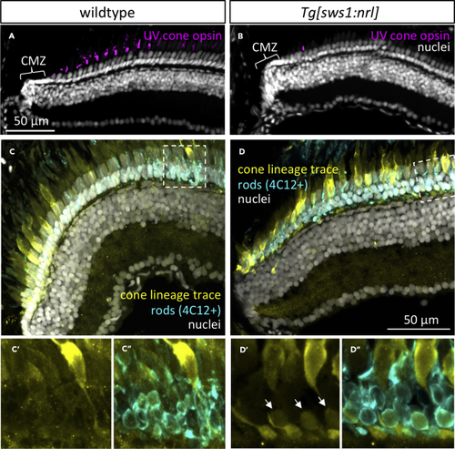
The Cone Cell Lineage Does Not Appreciably Contribute to Rod Production in Zebrafish but Inducing Ectopic Nrl Shows It has This Capacity

(A) Adult zebrafish retina grows from proliferating cells in the ciliary marginal zone (CMZ), generating all cell types including regularly spaced UV cones.

(B) Following ectopic expression of Nrl in differentiating UV cones, adult retina is mostly devoid of UV cones, except sparse newly born UV-opsin-positive cells near the CMZ. (A) and (B) are anti-UV opsin immunohistochemistry (magenta) with nuclear counterstain.

(C) The adult zebrafish cone cell lineage gives rise to all cone cell types, and few other cell types are appreciably generated from that lineage, as detected by Cre-lox lineage tracing via a cone-transducin-α (gnat2) Cre driver line. No history of gnat2 expression (yellow) is detectable in rod cells (4C12+), despite the lineage trace reporter being abundant in all other photoreceptors (cones). (C’) and (C″) are alternate views of dashed box outlined in (C). See also associated Figure S4.

(D) The fate of UV-opsin-positive cells that are absent from mature retina in (B) includes their transmutation into rods. Note a subset of rod cells (arrows, identified as 4C12+ and with nuclei in the basal-most layer of the ONL) shows a history of gnat2 expression (yellow), indicating they were generated from the cone lineage.

Nrl is Dispensable for Rod Specification in Adult Zebrafish

(A) Monitoring for appearance of GFP-positive rods during ontogeny of nrl−/− larvae directed our attention to 11 days postfertilization (dpf), where rods are sporadically detectable, but varied between individuals and between eyes of the same individual. Wild-type (WT) eyes at top with abundant rods provided for context.

(B–D) Adult nrl−/− zebrafish possess a large abundance of rods indistinguishable from WT, such that GFP-positive rods are obvious in intact animals (B) or retinal cryosections (C). Adult nrl−/− retina showed normal distribution of rod outer segments (ros) apical of rod cell bodies in the outer nuclear layer (ONL, also in [D]). Inner nuclear layers (INLs) and retinal ganglion cell layers (RGCs) are overtly normal (quantified in Figures S5G and S5H).

(E) Immunolabelling with rod-specific 4C12 and anti-UV-opsin confirms presence of rods and normal UV cones, respectively, in adult
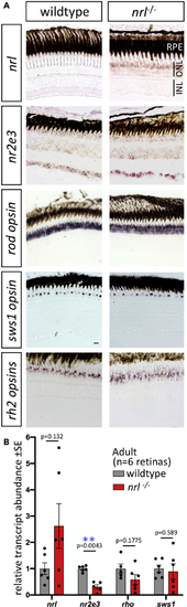
 nrl−/− retina.

Adult Retina of Zebrafish nrl−/− Mutants Show Changes in Abundance of Nrl Target Gene nr2e3 and in nrl Itself

(A) Gene expression determined by in situ hybridization on cryosections of adult zebrafish retina. Abundance of nrl transcript is higher in nrl−/− frameshift mutant retina (confirmed in panel B), suggesting an auto-regulatory negative feedback loop controlling it own abundance. Assuming that nrl transcript location is unaltered in nrl−/− mutants (despite aforementioned changes in its abundance), these data suggest expression of nrl is highly enriched in the outer nuclear layer, consistent with the site of phenotypes when Nrl protein is disrupted. Alterations to the abundance of nr2e3, a downstream target of Nrl, are equivocal when measured by in situ hybridization. The levels and distributions of transcripts encoding rod (rh1) and cone (sws1 and rh2) opsins were not detectably different in nrl−/− mutant retina compared to wild type. Scale bars represent 10 μm.

(B) Transcript abundance in adult neural retina determined by RT-qPCR confirms an increase in
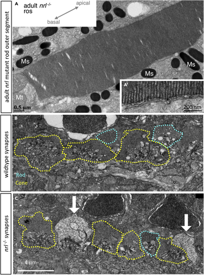
 nrl abundance in zebrafish bearing a frameshift null allele in nrl. A downstream transcriptional target of Nrl, nr2e3, was 70% reduced in abundance in nrl−/− mutant retina compared to wild type (p < 0.01; n = 5–6 individuals per genotype). Opsin abundances in adult retinas were not markedly different between genotypes.

Rod Outer Segments of nrl−/− Adult Zebrafish Appear Normal

(A) Rods demonstrate the expected hairpin end of floating disks within the outer segment that are non-contiguous with the outer cell membrane (A′), diagnostic of rod cell identity

(B) In wild-type adult zebrafish, photoreceptor synaptic terminals include rod spherules (teal dotted line) that are morphologically distinguishable from cone pedicles (yellow).

(C) In nrl−/− adult retina, the cone pedicles appear normal, and rod spherules appear normal but are very sparse; instead, electron-lucent terminals (white arrows) uniquely appear and may represent an Nrl-dependent defect in rod synapse maintenance. Various characters of photoreceptor terminals are quantified in Figure S6. Mt, mitochondria; Ms, melanosomes.

Acknowledgments
This image is the copyrighted work of the attributed author or publisher, and ZFIN has permission only to display this image to its users. Additional permissions should be obtained from the applicable author or publisher of the image. Full text @ iScience