FIGURE SUMMARY
Title

The novel zebrafish model pretzel demonstrates a central role for SH3PXD2B in defective collagen remodelling and fibrosis in Frank-Ter Haar syndrome

Authors
de Vos, I.J.H.M., Wong, A.S.W., Taslim, J., Ong, S.L.M., Syder, N.C., Goggi, J.L., Carney, T.J., van Steensel, M.A.M.
Source
Full text @ Biol. Open

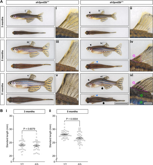
Adult sh3pxd2bΔ/Δ zebrafish have a gradually worsening phenotype, affecting skeletal structures and skin. (A) Representative images of sh3pxd2bΔ/Δ mutant fish and their sh3pxd2b+/+ WT clutch mates at the ages of 3, 5 and 11 months, selected from a total of 44 analysed fish per genotype per time point (see Table 1), imaged with a Nikon SMZ25 stereomicroscope. At 3 months of age, mutants demonstrate a short, up-tilted head (black arrowhead) with short operculum, exposing the gills (visible as a red patch caudal to the operculum); these features are absent in their WT clutch mates. At the age of 5 months, the pectoral fins of mutants are stiff and constantly kept in abducted position (blue arrowhead). At the anterior base of their dorsal fin, an opaque spot can be observed (iv, magenta arrow) that is absent in younger mutants and WT fish (compare with i–iii). At 11 months of age, mutants demonstrate hyper-kyphosis and abdominal constriction (black arrow). The pectoral fins of mutants are stiff and show signs of physical damage in multiple individuals (blue arrowhead). The opaque spot at the base of the dorsal fin (vi, magenta outlined arrow) is larger at 11 months of age and is hyper-pigmented (compare with iv and v). Note the sharper angle of fin rays with the dorsum of the fish in mutants (iv,vi) as compared to WT clutch mates (iii,v), as a result of the inability of the fins to be abducted manually. The flank, ventral to the dorsal fin, shows a whitish discoloration (green outlined arrows) in mutants. Scale bar equals 10 mm in overview and 1 mm in dorsal fin close-up. (B) Standard length measurements of 25 WT and 27 mutant 3-month-old fish (i), and 40 WT and 41 mutant 5-month-old fish. While there is no statistically significant difference in mean standard length between mutants and WT clutch mates at 3 months of age (i), at the age of 5 months mutants are significantly shorter than their WT clutch mates (P=0.6079 and P=0.0004, respectively, as assessed by two-sampled, non-pooled, two-tailed Student's t-test). Error bars represent standard error of means of biological replicates.

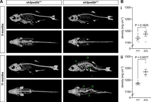
Adult sh3pxd2bΔ/Δ zebrafish develop kyphoscoliosis and an increased cutaneous density. (A) Representative µCT 3D renders of sh3pxd2bΔ/Δ mutant fish and sh3pxd2b+/+ WT clutch mates at the age of 5 and 11 months, selected form a total of five assessed fish per genotype per time point, imaged with a Siemens Inveon CT. At 5 months of age, mutant fish have a relatively small, up-tilted skull as compared to their WT clutch mates. At 11 months, mutants have developed kyphoscoliosis (outlined arrow). The skin of mutants shows an increased density (outlined arrow heads), that is not present at 5 months of age. Note that the fish displayed here had all the phenotypical features listed in Table 1 (see Fig. S2 for gross anatomy images). (B) Tissue density measurements of the five scanned fish per genotype per time point. At 5 months of age (i), there is a slight increase in overall tissue density of mutant (Δ/Δ) fish compared to their WT (+/+) clutch mates, which is however not statistically significant (NS; P=0.1625). At 11 months of age (ii), the overall tissue density of mutant fish is significantly increased compared to their WT clutch mates (P=0.0077; assessed by two-sampled, non-pooled, two-tailed Student's t-test). Error bars represent standard error of means of biological replicates.

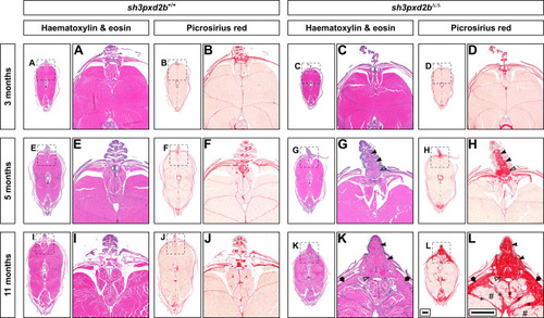
Adult sh3pxd2bΔ/Δ zebrafish develop cutaneous fibrosis that worsens over time. Representative H&E and PSR stained transverse sections at the level of the dorsal fin of sh3pxd2bΔ/Δ mutant fish and sh3pxd2b+/+ WT clutch mates at the ages of 3, 5 and 11 months, selected from five assessed fish per genotype per time point, imaged with a Zeiss AxioImager Z.2 slide scanner. At 3 months of age, transverse sections of mutants (C,D) and their WT clutch mates (A,B) are indistinguishable. At the age of 5 months, mutants have accumulated an increased amount of connective tissue at the base of the dorsal fin between the lepidotrichiae (G, solid arrow heads) and around the distal radial (outlined arrow head) as compared to their WT clutch mates (E), primarily consisting of collagen (F,H). At 11 months of age, the base of the dorsal fin in mutants is wider than their WT clutch mates and completely filled with collagen (I–L). In addition, collagen accumulates in the stratum compactum (arrows), myosepta (asterisks), and between individual muscle fibres (hash signs) in mutants. Scale bars equal 500 µm.

ZFIN is incorporating published figure images and captions as part of an ongoing project. Figures from some publications have not yet been curated, or are not available for display because of copyright restrictions.

EXPRESSION / LABELING:
Gene:
Fish:
Anatomical Term:
Stage: Days 45-89
Acknowledgments
This image is the copyrighted work of the attributed author or publisher, and ZFIN has permission only to display this image to its users. Additional permissions should be obtained from the applicable author or publisher of the image. Full text @ Biol. Open