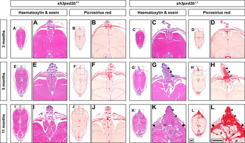
Adult sh3pxd2bΔ/Δ zebrafish develop cutaneous fibrosis that worsens over time. Representative H&E and PSR stained transverse sections at the level of the dorsal fin of sh3pxd2bΔ/Δ mutant fish and sh3pxd2b+/+ WT clutch mates at the ages of 3, 5 and 11 months, selected from five assessed fish per genotype per time point, imaged with a Zeiss AxioImager Z.2 slide scanner. At 3 months of age, transverse sections of mutants (C,D) and their WT clutch mates (A,B) are indistinguishable. At the age of 5 months, mutants have accumulated an increased amount of connective tissue at the base of the dorsal fin between the lepidotrichiae (G, solid arrow heads) and around the distal radial (outlined arrow head) as compared to their WT clutch mates (E), primarily consisting of collagen (F,H). At 11 months of age, the base of the dorsal fin in mutants is wider than their WT clutch mates and completely filled with collagen (I–L). In addition, collagen accumulates in the stratum compactum (arrows), myosepta (asterisks), and between individual muscle fibres (hash signs) in mutants. Scale bars equal 500 µm.
|
