Endothelial cilia are observed during early development of the hyaloid vasculature, but rapidly decline in number by 3 dpf.

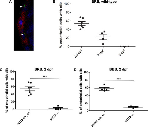
A) Confocal micrograph of the hyaloid vessels (red) of a 3 dpf Tg(flk1:mCherry;bactin2:Arl13b-GFP) embryo. Cilia (green, arrowheads) were observed on the endothelial cells, near the nuclei (DAPI, blue). B) Percentage of hyaloid vessel endothelial cells possessing a primary cilium at 2, 3, and 5 dpf (n > 80 endothelial cells from ≥ 4 eyes per time point). C) Percentage of hyaloid vessel endothelial cells possessing a primary cilium at 2 dpf in ift172 homozygous mutant fish compared to wild-type and heterozygous siblings (n ≥ 4 fish per group). D) Percentage of brain endothelial cells possessing a primary cilium at 2 dpf in ift172 homozygous mutant fish compared to wild-type and heterozygous siblings (n ≥ 3 fish per group). ***p < 0.0005 by Fisher’s exact test. Error bars = SEM.

IFT mutant fish develop and maintain a functional BRB.

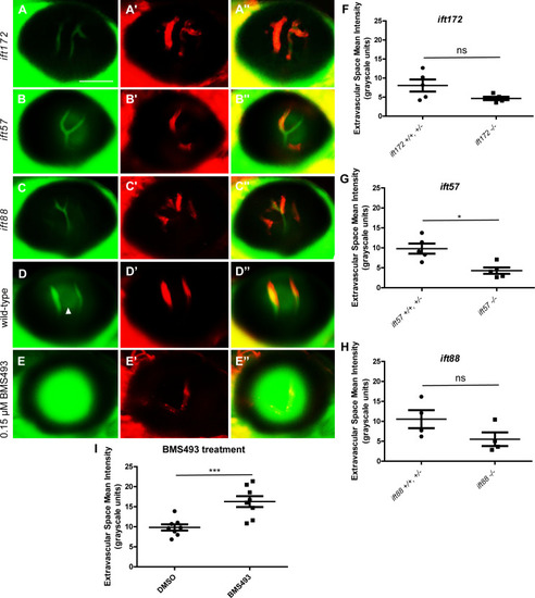
Fluorescent micrographs of the hyaloid vessels in live 6 dpf IFT mutant and wild-type Tg(l-fabp:DBP-EGFP;flk1:mCherry) fish. In the ift172 (A), ift57 (B), and ift88 (C) mutants and wild-type larvae (D), the DBP-EGFP- associated signal (green) localizes within the mCherry-tagged vessels (red), indicating that the BRB is intact. Some autofluorescence (likely from the lens) is observed in the areas outside of the hyaloid vessels (arrowhead in D). (E) As an example of BRB breakdown, Tg(l-fabp:DBP-EGFP;flk1:mCherry) embryos were treated with 0.15 μM BMS493, an inhibitor of retinoic acid signaling, from 2 dpf until 7 dpf. Scale bars = 50 μm. No increase in the Extravascular Space Mean Intensity was observed in the eyes of 6 dpf ift172 (F), ift57 (G), or ift88 (H) fish compared to their wild-type siblings, confirming that the IFT mutant fish do not exhibit increased leakage of DBP-EGFP outside of their hyaloid vessels. Interestingly, the Ift mutant fish actually trend towards a decreased level of autofluorescence in the extravasal space (* p < 0.05, ns = not significant, by Student’s t-test). To confirm the ability of this measurement technique to detect BRB leakage in larval fish, the Extravascular Space Mean Intensity of 7 dpf BMS493-treated fish was measured and compared to that of their DMSO (vehicle)-treated siblings (I) (*** p < 0.005, by Student’s t-test).

<italic>Ift</italic> mutant fish with ICH maintain an intact BRB.

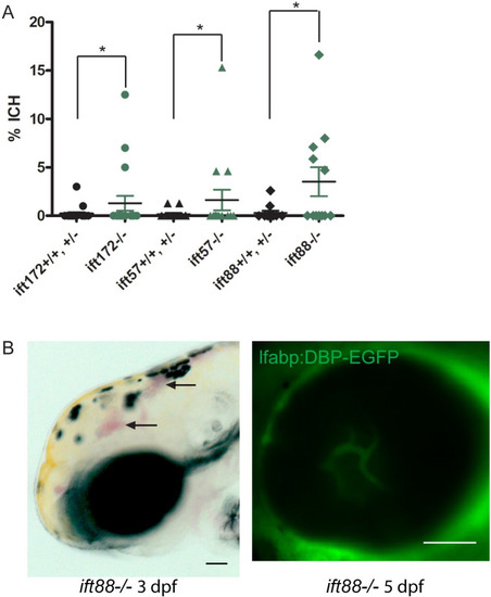
Embryos were screened daily for ICH and BRB integrity was assessed at 5 dpf. (A) Average percentages of ift172-/-, ift57-/-, and ift88-/- embryos and their wild type and heterozygous siblings with ICH (n = 18 ift172 crosses, 15 ift57 crosses, and 10 ift88 crosses of ≥ 50 embryos each). Error bars = SEM. *P < 0.05 by Fisher’s exact test. (B) An ift88-/- embryo with ICH at 3 dpf (bright field image at left, arrowheads) and an intact BRB at 5 dpf (confocal micrograph at right). Scale bar = 50 μm.

Early inhibition of the hedgehog signaling pathway results in severe edema and BRB non-perfusion.

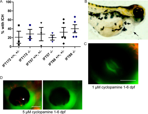
Zebrafish embryos were treated with 1 μM cyclopamine starting at 25 hpf. (A) Percentages of cyclopamine-treated IFT mutant and non-mutant embryos that developed ICH by 52 hpf. No significant differences were detected between mutants and non-mutants by Fisher’s exact test (p > 0.05). (B) Image of a 6 dpf wild-type fish that had cyclopamine-induced pericardial edema (arrow). (C) Fluorescent micrograph of the eye of a zebrafish treated with 1 μM cyclopamine from 1 to 6 dpf, demonstrating an intact blood-retinal barrier. (D) Fluorescent micrograph of a 5 μM cyclopamine-treated zebrafish at 6 dpf with non-perfusion of the hyaloid vasculature (arrowhead). Scale bars = 50 μm.

Later treatment of zebrafish larvae with higher doses of cyclopamine results in BRB leakage.

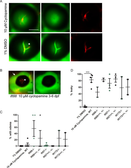
Zebrafish larvae were treated with 10 μM cyclopamine from 5 dpf– 7 dpf. (A) Cyclopamine-treated larvae had BRB breakdown at 7 dpf, as seen by the detection of DBP-EGFP (green) outside of the hyaloid vessels (red). Arrowhead indicates autofluorecence. (B) Fluorescent micrograph of a cyclopamine-treated zebrafish at 7 dpf with edema around the eye (arrow), and non-perfusion of the hyaloid vasculature (arrowhead). (C) Graph depicting the percentage of WT and Ift mutant fish in each clutch with edema/non-perfusion after treatment with 10 μM cyclopamine from 5 dpf– 7 dpf (differences between mutants and their non-mutant siblings not significant by Student’s t-test). (D) Graph depicting the percentage of WT and Ift mutant fish in clutches without edema/non-perfusion that had BRB breakdown upon treatment with 10 μM cyclopamine or 1% DMSO vehicle control (***p < 0.0005, all other differences not significant by Student’s t-test). Each data point in (C) and (D) represents a clutch of >50 larvae. Error bars represent SEM. Scale bars = 50 μm.

Acknowledgments
This image is the copyrighted work of the attributed author or publisher, and ZFIN has permission only to display this image to its users. Additional permissions should be obtained from the applicable author or publisher of the image. Full text @ PLoS One