IMAGE

Fig 1

ID
ZDB-IMAGE-200811-9
Source
Figures for Pollock et al., 2020
Image
Figure Caption

Fig 1 Endothelial cilia are observed during early development of the hyaloid vasculature, but rapidly decline in number by 3 dpf.

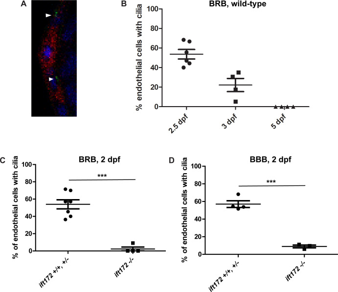
A) Confocal micrograph of the hyaloid vessels (red) of a 3 dpf Tg(flk1:mCherry;bactin2:Arl13b-GFP) embryo. Cilia (green, arrowheads) were observed on the endothelial cells, near the nuclei (DAPI, blue). B) Percentage of hyaloid vessel endothelial cells possessing a primary cilium at 2, 3, and 5 dpf (n > 80 endothelial cells from ≥ 4 eyes per time point). C) Percentage of hyaloid vessel endothelial cells possessing a primary cilium at 2 dpf in ift172 homozygous mutant fish compared to wild-type and heterozygous siblings (n ≥ 4 fish per group). D) Percentage of brain endothelial cells possessing a primary cilium at 2 dpf in ift172 homozygous mutant fish compared to wild-type and heterozygous siblings (n ≥ 3 fish per group). ***p < 0.0005 by Fisher’s exact test. Error bars = SEM.

Acknowledgments
This image is the copyrighted work of the attributed author or publisher, and ZFIN has permission only to display this image to its users. Additional permissions should be obtained from the applicable author or publisher of the image. Full text @ PLoS One