IMAGE

Fig 2

ID
ZDB-IMAGE-200811-10
Source
Figures for Pollock et al., 2020
Image
Figure Caption

Fig 2 IFT mutant fish develop and maintain a functional BRB.

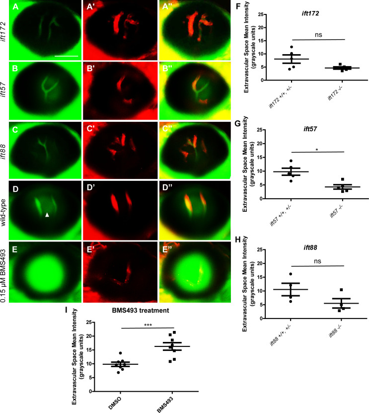
Fluorescent micrographs of the hyaloid vessels in live 6 dpf IFT mutant and wild-type Tg(l-fabp:DBP-EGFP;flk1:mCherry) fish. In the ift172 (A), ift57 (B), and ift88 (C) mutants and wild-type larvae (D), the DBP-EGFP- associated signal (green) localizes within the mCherry-tagged vessels (red), indicating that the BRB is intact. Some autofluorescence (likely from the lens) is observed in the areas outside of the hyaloid vessels (arrowhead in D). (E) As an example of BRB breakdown, Tg(l-fabp:DBP-EGFP;flk1:mCherry) embryos were treated with 0.15 μM BMS493, an inhibitor of retinoic acid signaling, from 2 dpf until 7 dpf. Scale bars = 50 μm. No increase in the Extravascular Space Mean Intensity was observed in the eyes of 6 dpf ift172 (F), ift57 (G), or ift88 (H) fish compared to their wild-type siblings, confirming that the IFT mutant fish do not exhibit increased leakage of DBP-EGFP outside of their hyaloid vessels. Interestingly, the Ift mutant fish actually trend towards a decreased level of autofluorescence in the extravasal space (* p < 0.05, ns = not significant, by Student’s t-test). To confirm the ability of this measurement technique to detect BRB leakage in larval fish, the Extravascular Space Mean Intensity of 7 dpf BMS493-treated fish was measured and compared to that of their DMSO (vehicle)-treated siblings (I) (*** p < 0.005, by Student’s t-test).

Acknowledgments
This image is the copyrighted work of the attributed author or publisher, and ZFIN has permission only to display this image to its users. Additional permissions should be obtained from the applicable author or publisher of the image. Full text @ PLoS One