FIGURE SUMMARY
Title

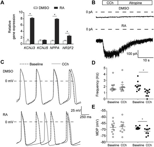
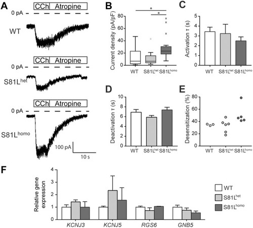
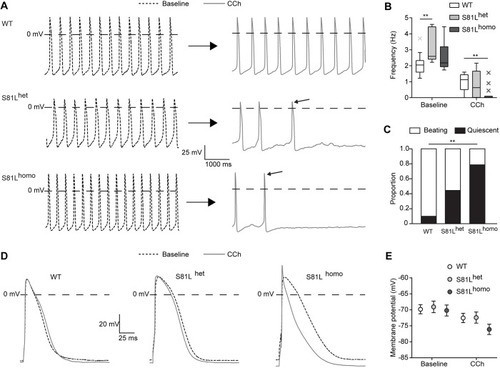
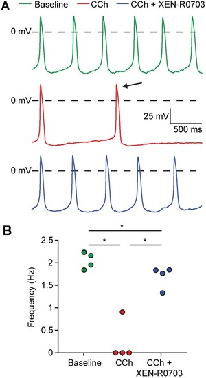
Genetic variation in GNB5 causes bradycardia by augmenting the cholinergic response via increased acetylcholine-activated potassium current (IK,ACh)

Authors
Veerman, C.C., Mengarelli, I., Koopman, C.D., Wilders, R., van Amersfoorth, S.C., Bakker, D., Wolswinkel, R., Hababa, M., de Boer, T.P., Guan, K., Milnes, J., Lodder, E.M., Bakkers, J., Verkerk, A.O., Bezzina, C.R.
Source
Full text @ Dis. Model. Mech.

Application of retinoic acid (RA) to differentiating hiPSCs results in cardiomyocytes (CMs) that exhibit a robust acetylcholine-activated K+ current (IK,ACh). (A) Transcript expression of genes in RA-treated hiPSC-CMs relative to expression in DMSO-treated hiPSC-CMs. Data are mean±s.e.m. of three biological replicates, normalized to TBP. RA treatment enhanced expression of the genes KCNJ3, NPPA and NR2F2. *P<0.05 (two-sided t-tests). (B) Typical IK,ACh traces in DMSO- and RA-treated hiPSC-CMs initiated by fast application of 100 µmol l−1 carbachol (CCh). After reaching steady state, addition of 1 mmol l−1 atropine achieves instant removal of CCh from muscarinic receptors and deactivation of the current. In DMSO-treated hiPSC-CMs, IK,ACh was not detected, while RA-treated hiPSC-CMs all demonstrated a robust current (for average data, see Fig. 2). (C) Typical spontaneous action potentials (APs) of DMSO- and RA-treated hiPSC-CMs at baseline and upon addition of 10 µmol l−1 CCh. (D,E) Frequency of spontaneous APs (D: n=9, DMSO; n=10, RA) and maximal diastolic potential (MDP; E: n=9, DMSO; n=9, RA) of each cell at baseline and in the presence of CCh in DMSO- and RA-treated hiPSC-CMs. *P<0.05 (two-way ANOVA). CCh reduces the AP frequency and causes significant MDP hyperpolarization in RA-treated, but not in DMSO-treated, hiPSC-CMs.

Impact of the Gβ5-S81L variant on IK,ACh in RA-treated hiPSC-CMs. (A,B) Typical recordings (A) and average IK,ACh density (B) in WT, S81Lhet and S81Lhomo hiPSC-CMs. Average IK,ACh density is increased in S81Lhomo hiPSC-CMs compared to WT and S81Lhet hiPSC-CMs. *P<0.05 (Kruskal–Wallis non-parametric test, followed by Bonferroni post-hoc analysis). (C,D) Time constant (τ) of IK,ACh activation (C) and deactivation (D). (B-D) WT, n=27; S81Lhet, n=11; S81Lhomo, n=20 hiPSC-CMs. (E) Percentage of desensitization, measured as the percentage of IK,ACh decrease at steady state upon continuous CCh application; WT, n=3; S81Lhet, n=6; S81Lhomo, n=5 hiPSC-CMs. (F) Transcript level of the genes involved in G-protein-coupled activation of IK,ACh. Data are mean±s.e.m. of three biological differentiation replicas and expression level of each gene in S81Lhet and S81Lhomo hiPSC-CMs is indicated as relative to its expression in WT hiPSC-CMs. No significant differences are observed in KCNJ3, KCNJ5, RGS6 and GNB5 transcript abundance across the WT, S81Lhet and S81Lhomo hiPSC-CMs.

CCh induces more pronounced effects in S81Lhomo compared to WT and S81Lhet RA-treated hiPSC-CMs. (A) Representative spontaneous APs at baseline (left) and upon 10 µmol l−1 CCh (right) in WT, S81Lhet and S81Lhomo RA-treated hiPSC-CMs. The slanted arrows near the APs in the presence of CCh indicate the last AP before cessation of spontaneous activity. (B) Average frequencies before and during application of CCh. CCh drastically reduces the spontaneous AP frequency of S81Lhomo hiPSC-CMs. WT, n=10; S81Lhet, n=9; S81Lhomo, n=14. **P<0.01 (two-way repeated measures ANOVA and post-hoc Bonferroni corrected pairwise comparison). (C) Proportion of WT, S81Lhet and S81Lhomo hiPSC-CMs that became quiescent upon application of CCh. WT, n=10; S81Lhet, n=9; S81Lhomo, n=14. **P<0.01 (chi-square test, followed by Bonferroni corrected pairwise comparisons). (D) Representative APs (1 Hz overdrive stimulation) before and after the application of CCh. (E) Average maximal diastolic potential (MDP) at baseline and upon CCh in WT, S81Lhet and S81Lhomo RA-treated hiPSC-CMs (1 Hz overdrive stimulation). WT, n=17; S81Lhet, n=10; S81Lhomo, n=13. Effect of CCh on MDP is significantly more pronounced in S81Lhomo hiPSC-CMs (P<0.05 interaction effect CCh×genotype; two-way ANOVA).

CCh induces an extreme slowing of spontaneous beating in S81Lhomo hiPSC-CMs, which is largely suppressed by IK,ACh blockade through XEN-R0703. (A) Typical APs of an S81Lhomo hiPSC-CM at baseline (top), upon application of 10 µmol l−1 CCh (middle), and upon addition of 1 µmol l−1 XEN-R0703 in the presence of 10 µmol l−1 CCh (bottom). (B) Effects of CCh and XEN-R0703 on spontaneous AP frequency in four S81Lhomo hiPSC-CMs. *P<0.05 (one-way repeated measures ANOVA followed by post-hoc Holm-Sidak test). XEN-R0703 restores the beating frequency of S81Lhomo hiPSC-CMs to values similar to a baseline condition.

Effect of IK,ACh blockade on heart rate (HR) in gnb5-knockout zebrafish. (A) Experimental setup for high-speed imaging of the zebrafish heart. HR was recorded at 150 fps for 10 s. (B) Time schedule of drug treatment and CCh challenge in gnb5-knockout larvae. (C) Scatter dot plots with mean of HR of gnb5-knockout larvae 5 days post-fertilization at basal level, after treatment with 50 µmol l−1 XEN-R0703 or DMSO, and upon application of 500 µmol l−1 CCh. DMSO, n=13; XEN-R0703, n=22. **P<0.01, ***P<0.001, ****P<0.0001 (two-way repeated measures ANOVA followed by Bonferroni corrected pairwise comparisons). (D) Percentage of gnb5-knockout larvae that responded to 500 µmol l−1 CCh with a decrease in HR of 0-25, 26-50, 51-75 or 76-100%. DMSO, n=13; XEN-R0703, n=22. Only 27% of gnb5-knockout larvae pretreated with XEN-R0703 displayed a pathological decrease in HR (51-75%, 76-100%), while over 80% of the not-pretreated gnb5-knockout larvae display the same decreased level of HR.

ACh induces more pronounced effects in S81Lhomo compared to WThuman SAN pacemaker cells in computer simulations. (A-C) Spontaneous APs (blue lines) and associated net membrane current (Inet, gray lines), hyperpolarization-activated ‘funny current’ (If, green lines) and IK,ACh (red lines) in a WT human SAN pacemaker cell at baseline (A), upon addition of 10 nmol l−1 ACh (B) and upon addition of 10 nmol l−1 ACh in the presence of XEN-R0703 (C). (D-F) Spontaneous APs and associated Inet, If and IK,ACh in an S81Lhomo human SAN pacemaker cell at baseline (D), upon addition of 10 nmol l−1 ACh (E) and upon addition of 10 nmol l−1 ACh in the presence of XEN-R0703 (F).

ACh induces more pronounced AP effects in S81Lhomo compared to WT human atrial myocytes in computer simulations. (A,B) APs elicited at 1 Hz (top panels) and associated IK,ACh (bottom panels) at baseline (dotted lines) and upon addition of 10 nmol l−1 ACh (solid lines) in WT (A) and S81Lhomo (B) human atrial myocytes. The horizontal double-headed arrow in panel A indicates the ACh-induced shortening of AP duration. The ‘wobbles’ in the time course of IK,ACh in panel B are caused by the N-shape of the IK,ACh current-voltage relationship (insets). The slanted arrows in the insets point to the IK,ACh reversal potential (EK) of −83.0 mV. (C,D) Shift in maximum diastolic potential (ΔMDP; C) and threshold stimulus current amplitude (D) at ACh concentrations ranging from 0.1 nmol l−1 to 1 µmol l−1. Note the logarithmic abscissa scale.

Acknowledgments
This image is the copyrighted work of the attributed author or publisher, and ZFIN has permission only to display this image to its users. Additional permissions should be obtained from the applicable author or publisher of the image. Full text @ Dis. Model. Mech.